
Page 548 - IJB-10-4
P. 548
International Journal of Bioprinting 3D-bioprinting of osteochondral plugs
the chondrogenic medium (Figure 6E–H). As some cells 3.6. OsteoImage assay
are stained positively for both live and dead markers, Human bone marrow-derived mesenchymal stem cells
quantifying viability becomes challenging. Additionally, (hbMSCs) embedded in the bone bioink described in
hbMSCs were also seeded on 3D-printed PCL disks. Cells Section 3.5 were evaluated for their ability to undergo
attached to the material proliferated and maintained high nascent bone formation. Following 28 days of culture in
viability over 14 days (Figure S1, Supporting Information). either osteogenic or growth media, bone bioink disks were
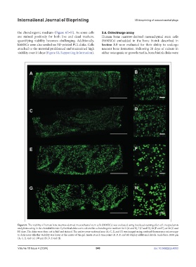
Figure 6. The viability of human bone marrow-derived mesenchymal stem cells (hbMSCs) was evaluated using live/dead staining after cell encapsulation
and photocuring in the chondral bioink. Cylindrical disks were cultured in a chondrogenic medium for 1 (A and B), 7 (C and D), 28 (E and F), or 56 (G and
H) days. The disks were then cut in half and stained. The entire cross-sectional area (A, C, E, and G) was imaged using confocal fluorescence microscopy
to determine whether viability was lower at the center of the gel. Insets at each time point (B, D, F, and H) display additional details. Scale bars: 1000 µm
(A, C, E, and G); 100 µm (B, D, F, and H).
Volume 10 Issue 4 (2024) 540 doi: 10.36922/ijb.4053