
Page 108 - IJB-10-5
P. 108
International Journal of Bioprinting 3D bioprinting for organoid-derived EVs
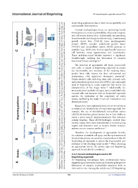
extracellular matrix (dECM), and biomolecules including signaling molecules. (B) Four main 3D printing methods are inkjet bioprinting, laser-assisted bioprinting, extrusion bioprinting, and
bioprinting applications due to their biocompatibility and
Figure 2. Overview of bioink composition, common bioprinting methods, and their applications. (A) The main elements of bioinks are cells, support materials like hydrogel, polymers like decellularized
photocuring bioprinting. (C) The applications of 3D bioprinting range from tissue construct printing and disease model establishment to drug testing and delivery. Schematic created with BioRender.
customizable characteristics.
Current methodologies focus on optimizing bioink
formulations to enhance printability, structural integrity,
and cell-matrix interactions. Additionally, incorporating
bioactive molecules like growth factors (e.g., transforming
growth factor beta [TGF-β], bone morphogenetic
protein [BMP], vascular endothelial growth factor
[VEGF]) and extracellular matrix (ECM) proteins or
peptides (e.g., RGD) into bioinks significantly improves
cell behavior, tissue regeneration, and functionality.
These multifunctional bioinks represent a significant
breakthrough, enabling the fabrication of complex
functional tissues and organs. 32
The selection of appropriate cell types, particularly
stem cells, is crucial in bioprinting organoids to ensure
the functionality and relevance of the resulting tissue
model. Stem cells, known for their self-renewal and
pluripotency, offer significant therapeutic potential.
33
Patient-derived cells, including stem cells, primary cells,
and induced pluripotent stem cells (iPSCs), are commonly
used in bioprinting to create organoids that mimic the
34
characteristics of the target tissue. Additionally, the
incorporation of multiple cell types, such as epithelial cells,
stromal cells, and immune cells, in bioprinted organoids
enables the replication of the complexity of native
tissues, facilitating the study of cell-cell interactions and
disease processes.
Researchers have explored diverse bioink formulations
to enhance the functionality of bioprinted organoids. For
example, the use of decellularized extracellular matrix
(dECM) bioinks has gained interest due to their ability to
create a more natural microenvironment that enhances
cellular function. These dECM hydrogels, derived from
various tissues, have been instrumental in reconstructing
organs and structures such as tumors, hearts, muscles,
arteries, nerves, corneas, and bones. 35
Therefore, the development of appropriate bioinks,
the selection of suitable cell types, and the optimization of
bioink formulations are essential considerations in the 3D
bioprinting of organoids to ensure the functionality and
relevance of the resulting tissue model. When choosing
36
an appropriate bioink, the printer type and bioprinting
approach should also be considered.
2.2.2. Current methodologies used in 3D
bioprinting organoids
3D bioprinting technologies have revolutionized tissue
engineering and precision medicine by enabling the precise
fabrication of complex multicellular structures both in
vitro and in vivo. Among the widely used 3D bioprinting
Volume 10 Issue 5 (2024) 100 doi: 10.36922/ijb.4054