
Page 107 - IJB-10-5
P. 107
International Journal of Bioprinting 3D bioprinting for organoid-derived EVs
the personalization of treatment strategies based on the
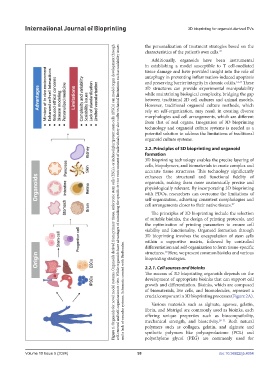
self-renewal and self-organization in vitro. While organoids have advantages of mimicking the specific in vivo environment of individuals, they also suffer technical limitations such as scalability issues
Figure 1. Organoids for novel model systems. Organoids derived from primary tissue, embryonic stem cells (ESCs), or induced pluripotent stem cells (iPSCs) can model organ development through
characteristics of the patient’s own cells. 26
Additionally, organoids have been instrumental
in establishing a model susceptible to T cell-mediated
tissue damage and have provided insight into the role of
autophagy in preventing inflammation-induced apoptosis
and preserving barrier integrity in chronic colitis. 12,13 These
3D structures can provide experimental manipulability
while maintaining biological complexity, bridging the gap
between traditional 2D cell cultures and animal models.
However, traditional organoid culture methods, which
rely on self-organization, may result in creating diverse
morphologies and cell arrangements, which are different
from that of real organs. Integration of 3D bioprinting
technology and organoid culture systems is needed as a
potential solution to address the limitations of traditional
organoid culture systems.
2.2. Principles of 3D bioprinting and organoid
formation
3D bioprinting technology enables the precise layering of
cells, biopolymers, and biomaterials to create complex and
accurate tissue structures. This technology significantly
enhances the structural and functional fidelity of
organoids, making them more anatomically precise and
physiologically relevant. By incorporating 3D bioprinting
with PDOs, researchers can overcome the limitations of
self-organization, achieving consistent morphologies and
cell arrangements closer to their native tissues. 27
The principles of 3D bioprinting include the selection
of suitable bioinks, the design of printing protocols, and
the optimization of printing parameters to ensure cell
viability and functionality. Organoid formation through
3D bioprinting involves the encapsulation of stem cells
within a supportive matrix, followed by controlled
and a lack of vascular systems. Schematic created with BioRender.
differentiation and self-organization to form tissue-specific
10
structures. Here, we present common bioinks and various
bioprinting strategies.
2.2.1. Cell sources and bioinks
The success of 3D bioprinting organoids depends on the
development of appropriate bioinks that can support cell
growth and differentiation. Bioinks, which are composed
of biomaterials, live cells, and biomolecules, represent a
crucial component in 3D bioprinting processes (Figure 2A).
Various materials such as alginate, agarose, gelatin,
fibrin, and Matrigel are commonly used as bioinks, each
offering unique properties such as biocompatibility,
mechanical strength, and bioactivity. 28–31 Both natural
polymers such as collagen, gelatin, and alginate and
synthetic polymers like polycaprolactone (PCL) and
polyethylene glycol (PEG) are commonly used for
Volume 10 Issue 5 (2024) 99 doi: 10.36922/ijb.4054