
Page 140 - IJB-10-5
P. 140
International Journal of Bioprinting Liver printing: from structure to application
vivo functionality. However, primary hepatocytes exhibit pluripotent stem cells. Nonetheless, current challenges
reduced proliferation capacity once removed from the in stem cell differentiation include low differentiation
in vivo microenvironment, and they begin to experience efficiency, long differentiation time, and the production of
oxidative stress, leading to a rapid decline in function, heterogeneous cell populations. Subsequent research has
making them unsuitable for passaging and large-scale found that mimicking the liver microenvironment and co-
expansion. HepG2 liver cancer cell lines have the ability for culturing hepatocytes with other cell types can enhance
indefinite passaging, but they still exhibit some functional the maintenance of hepatocyte function. For instance,
differences compared to primary cells. HepaRG cells are hepatic stellate cells can mimic the ECM environment
bipotent liver progenitor cells with high proliferative of the liver; mesenchymal stem cells can enhance liver
potential, capable of differentiating into cholangiocyte- function via paracrine secretion of cytokines; endothelial
and hepatocyte-like cells. HepaRG cells, after dimethyl cells can accelerate vascularization and provide oxygen
sulfoxide (DMSO)-induced differentiation, can express and nutrients required by hepatocytes; and cholangiocytes
most major cytochrome P450 (CYP), such as CYP3A4, can promote bile duct system formation and liver-specific
2E1, and 1A2, making them a powerful tool for studying gene expression. 142,143 Furthermore, 3D hepatocyte
drug metabolism and hepatotoxicity. However, HepaRG cultures, such as hepatocyte spheroids and liver organoids,
cells still exhibit significant gaps in other liver functions, can induce polarity in hepatocytes, forming a closely
such as ALB production, urea synthesis, and bile secretion, connected spatial structure. This environment supports
compared to primary hepatocytes. Additionally, their long the formation of bile duct structures, which are crucial for
differentiation period (approximately 28 days) limits their bile production by hepatocytes. 144–147
broader use. 133–137
4.3. Inkjet bioprinting
Stem cells have the potential to differentiate into The selection of printing method generally depends on
various cell types and provide a regenerative source the type of bioink used and the desired printing accuracy
of hepatocytes, such as ESCs and iPSCs. Pluripotent and structural complexity (Figure 6B). Table 3 compares
21
stem cells initiate differentiation from the inner cell the different bioprinting technologies. Inkjet-based 3D
mass and undergo directed differentiation to become bioprinting is the earliest form of bioprinting, sharing
hepatic progenitor cells. These hepatic progenitor cells similarities with traditional inkjet printing. It utilizes
can subsequently differentiate into both hepatocytes and piezoelectric or thermal-driven printheads to continuously
cholangiocytes. 84,139–142 Additionally, non-parenchymal deposit tiny droplets of bioink onto a substrate, and the layer-
cells, such as liver sinusoidal endothelial cells, hepatic by-layer printing approach forms cell-laden 3D structures.
stellate cells, and Kupffer cells, can also be derived from Inkjet printing boasts high speed and resolution (up to 50
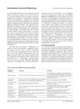
Table 3. Comparison of different bioprinting techniques
Bioprinting Advantages Limitations
technique
Low driving pressure, challenging to print high-viscosity
(>10 cP) materials and high-density cells; weak mechanical
Inkjet bioprinting Fast printing speed; high resolution (50 μm); relatively low-cost
integrity; uneven droplet size; potential for mechanical or
thermal damage to cells
Laser-assisted No nozzle, eliminating nozzle clogging and mechanical damage High cost; long printing time; low cell viability
bioprinting to cells; high precision (10 μm); supports high cell density
Photocuring-based Fast printing speed; high resolution (<100 μm) Applicable only to photocrosslinkable bioinks; requires
bioprinting long post-processing time
Low printing resolution (30–500 μm); does not support
Volumetric Fast printing speed (<1 min); supports large-volume structure multi-material, multi-cellular printing; low light
bioprinting printing (cm scale)
penetration
Extrusion-based Supports printing of high-viscosity materials and high-density Slow printing speed; low resolution (>100 μm); cell
bioprinting cell solutions viability may be reduced due to shear forces
Suspension-based Supports extrusion-based printing of low-viscosity materials; Complex post-processing; uneven material deposition;
bioprinting high printing accuracy (<50 μm) slow printing speed
Coaxial printing Coaxial multi-material, multi-cell printing Does not support printing branched structures
Volume 10 Issue 5 (2024) 132 doi: 10.36922/ijb.3819