
Page 143 - IJB-10-5
P. 143
International Journal of Bioprinting Liver printing: from structure to application
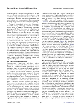
Generally, photocuring-based printing does not require carotid artery and jugular vein. Kang et al. employed a
160
extrusion through a nozzle, thus offering faster printing preset extrusion-based bioprinting technique to create liver
speeds and a higher resolution (<100 μm). However, its lobule structures containing multiple cells and materials.
applicability is limited to light-crosslinkable bioinks and These biomimetic liver lobules included hepatocytes,
requires longer post-processing times, thereby restricting endothelial cells, and complex tubular structures
the range of applications for photocuring-based printing. (Figure 7F). The preset models exhibited excellent structural
integrity and higher levels of ALB and urea synthesis, as well
Volumetric bioprinting is an innovative photocuring-
based 3D printing technology. Unlike SLA and DLP, as expression levels of ALB, MRP2, and CD31, compared
to co-cultures of hepatocytes and endothelial cells
which build up materials layer-by-layer, volumetric alone (Figure 7G). Similarly, Hong et al. utilized preset
27
printing rapidly forms 3D objects in a liquid vat by extrusion-based bioprinting combined with a microfluidic
projecting repeated light images from different angles emulsification technique to produce 3D-vascularized liver
into a transparent photopolymer liquid. This method tissue spheroids. These liver tissue spheroids featured liver
can complete printing within minutes (or even seconds) lobule-like cross-sections, complete tissue structures, and
without generating harmful mechanical stress on cells. high levels of MRP2, ALB, and CD31 expression. Yang
157
99
Bernal et al. used volumetric printing technology to et al. used gelatin and alginate-encapsulated HepaRG cells
construct complex 3D structures containing liver organoids as bioink to construct 3D-bioprinted liver-like organs
at centimeter-scale within 20 s. They selected 5% GelMA via extrusion-based printing (Figure 7H). Increasing
as the bioresin and added LAP as the photoinitiator. By the concentration of DMSO added during the induction
introducing iodixanol, they adjusted the optical properties process facilitated cholangiocyte differentiation (Figure
of the bioresin to address cell-mediated scattering issues. 7I). Protein expression of ALB, MRP2, and CYP3A4
The volumetric bioprinted liver organoids exhibited good in HepaRG cells within 3D-bioprinted hepatorganoids
cell viability, retaining their structure and polarity. (3DP-HOs) was significantly higher than in 2D cultures.
158
Currently, volumetric bioprinting faces limitations such Additionally, levels of ALB, α-1-antitrypsin, and factor VII
as low printing resolution (30–500 μm), which makes it secreted by 3DP-HOs were comparable to those of primary
more suitable for constructing centimeter-scale structures. human hepatocytes. Currently, extrusion-based printing
161
Additionally, improving light penetration and achieving faces challenges with slow printing speeds, low resolution
multi-material, multi-cellular volumetric printing are (>100 μm), and reduced cell viability due to shear force.
urgent challenges that need to be addressed.
4.7. Suspension-based bioprinting
4.6. Extrusion-based bioprinting High-viscosity bioinks are often selected to ensure high
Extrusion-based bioprinting technology utilizes fidelity and stable deposition, which limits the selection
pneumatic- or mechanically-driven nozzles to extrude range of bioinks. Additionally, high-viscosity materials
bioink in a controlled manner, depositing it onto a often require high extrusion pressure for deposition,
platform to form a 2D structure. As the nozzle or platform leading to decreased cell viability. In this regard,
moves in the z-axis direction, layers are stacked to create suspension-based bioprinting allows for the use of low-
3D structures. Extrusion-based bioprinting technology viscosity materials, such as collagen, fibronectin, and
evolved from inkjet printing and is currently the most cells without biomaterials. This technique also utilizes
widely used bioprinting technique due to its ability to extrusion-based 3D printers, but does not deposit
handle high-viscosity biological materials and high-density materials downwards onto flat surfaces; instead, it deposits
seed cells, utilizing a wide range of available materials. For them into a supporting bath of suspension medium,
instance, Miller et al. constructed a rigid filament network preventing the settling and collapse of printed structures.
using carbohydrate glass via extrusion-based printing. Suspension media have unique mechanical properties;
These networks, serving as cell-compatible sacrificial they exhibit solid-like characteristics under little or no
templates, generated hollow cylindrical networks that can stress, transitioning to liquid-like properties after applying
be lined with endothelial cells and perfused with blood, stress greater than the yield stress. When the bioink enters
making them an ideal strategy for constructing vascularized the suspension medium, microstructures spontaneously
tissues. Liu et al. designed a multi-material bioprinter recover, transforming the medium from liquid to solid
159
with multi-level temperature control based on extrusion- state and encapsulating the deposited material. The
162
based 3D printing to construct a centimeter-scale 3D liver Feinberg team printed collagen into a suspension bath
tissue model containing branched vascular networks. based on gelatin microspheres to manufacture trileaflet
The vascularized liver tissue facilitated the formation of heart valves. This Freeform Reversible Embedding of
capillary networks and direct surgical anastomosis of the Suspended Hydrogels (FRESH) technique reported a
Volume 10 Issue 5 (2024) 135 doi: 10.36922/ijb.3819