
Page 148 - IJB-10-5
P. 148
International Journal of Bioprinting Liver printing: from structure to application
emulsifying fats, transporting detoxification products there is still limited work combining liver tissue with bile
from the liver, and promoting the absorption of fatty duct structures, which holds significant importance. For
acids and fat-soluble vitamins. From a developmental example, co-culturing hepatocytes and cholangiocytes
41
biology perspective, intrahepatic bile ducts originate from introduces bile duct structures into liver organoids or
the hepatic region of the ventral foregut endoderm, while hepatocyte spheroids. These cells can be (i) primary
extrahepatic bile ducts emerge from the pancreatic ductal hepatocytes and primary cholangiocytes; or (ii) hepatocytes
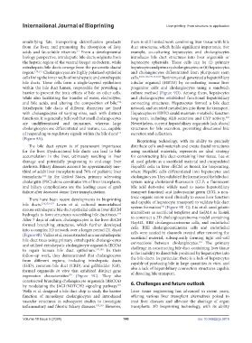
region. 170,171 Cholangiocytes are highly polarized epithelial and cholangiocytes differentiated from pluripotent stem
cells lining the inner walls of intrahepatic and extrahepatic cells. 82,84,138,139,144,183 Tanimizu et al. generated a hepatobiliary
bile ducts. These cells form a single-layered epithelium tubular organoid (HBTO) by co-culturing mouse liver
within the bile duct lumen, responsible for providing a progenitor cells and cholangiocytes using a sandwich
barrier to prevent the toxic effects of bile on other cells, culture method (Figure 9D). Among them, hepatocytes
while also facilitating the transfer of water, electrolytes, and cholangiocytes established functional hepatobiliary
and bile acids, and altering the composition of bile. connecting structures. Hepatocytes formed a bile duct
172
Intrahepatic bile ducts of different diameters are lined network and secreted metabolites into them for transport.
with cholangiocytes of varying sizes, each with distinct Hepatocytes in HBTO could maintain metabolic function
functions. It is generally believed that small cholangiocytes long-term, including ALB secretion and CYP activity.
183
are undifferentiated and immature, whereas large Nevertheless, current hepatobiliary organoids lack ductal
cholangiocytes are differentiated and mature, i.e., capable structures for bile excretion, preventing directional bile
of responding to regulatory signals within the bile ducts excretion and collection.
173
(Figure 9A). Bioprinting technology, with its ability to precisely
The bile duct system is of paramount importance distribute cells and materials and create ductal structures
for the liver. Dysfunctional bile ducts can lead to bile using sacrificial materials, represents an ideal strategy
accumulation in the liver, ultimately resulting in liver for constructing bile duct-containing liver tissue. Lee et
damage and potentially progressing to end-stage liver al. used gelatin as a sacrificial material and encapsulated
cirrhosis. Biliary diseases account for approximately one- HepaRG cells in liver dECM as bioink for bioprinting,
third of adult liver transplants and 70% of pediatric liver where HepaRG cells differentiated into hepatocytes and
transplants. In the United States, primary sclerosing cholangiocytes. They validated the formation of the bile duct
174
cholangitis (PSC) alone constitutes 5% of liver transplants, system using cholesterol-fluorescein (CLF; a fluorescent
and biliary complications are the leading cause of graft bile acid derivative widely used to assess hepatobiliary
failure after deceased donor liver transplantation. transport function) and indocyanine green (ICG; a non-
toxic organic anion used clinically to assess liver function
There have been recent developments in bioprinting
bile ducts. 174,175–177 Lewis et al. cultured immortalized and capable of hepatocyte transport) to validate bile duct
system formation (Figure 9E–G). Liu et al. used gelatin
142
mouse extrahepatic bile duct epithelial cells in liver dECM microfibers as sacrificial templates and GelMA as bioink
hydrogels to form structures resembling bile duct trees. to construct a 3D cholangiocarcinoma model containing
177
After 7 days of culture, cholangiocytes in the liver dECM HepG2, RBE cholangiocarcinoma cells, and endothelial
formed branching structures, which further developed cells. RBE cholangiocarcinoma cells and endothelial
into a complex 3D network over a longer period (21 days) cells were seeded in channels created after removing the
(Figure 9B). Vallier et al. reconstructed murine extrahepatic sacrificial material, subsequently forming tight cell–cell
bile duct trees using primary extrahepatic cholangiocytes connections between cholangiocytes. The primary
143
and utilized extrahepatic cholangiocyte organoids (ECOs) challenge in constructing bile duct-containing liver tissue
to repair human bile duct epithelium. 179,180 In their is the inability to direct bile produced by hepatocytes into
follow-up work, they demonstrated that cholangiocytes the bile ducts. In particular, there is a lack of hepatocytes
from different regions, including intrahepatic ducts capable of producing bile in large quantities in vitro, and
(IHD), common bile duct (CBD), and gallbladder (GB), also a lack of hepatobiliary connection structures capable
formed organoids in vitro that exhibited distinct gene of directing bile transport.
expression characteristics (Figure 9C). They also
179
constructed branching cholangiocyte organoids (BRCOs) 6. Challenges and future outlook
by modulating the JAG1/NOTCH2 signaling pathway.
181
Wells et al. designed a bile duct chip to study the barrier Liver tissue engineering has advanced in recent years,
function of monolayer cholangiocytes and introduced offering various liver transplant alternatives poised to
vascular structures in subsequent studies to investigate treat liver diseases and alleviate the shortage of organ
inflammatory and fibrotic biliary diseases. 182,183 However, transplants. 3D bioprinting technology, with its ability
Volume 10 Issue 5 (2024) 140 doi: 10.36922/ijb.3819