
Page 253 - IJB-10-5
P. 253
International Journal of Bioprinting 3D printed hydrogels for tumor therapy
Figure 8C–E graphically represents the changes of be similar. The pH of GelMA, HAp/GelMA, MgHAp/
pH value, Ca , and Mg in PBS after soaking GelMA, GelMA, and MgHAp/GelMA-PDA hydrogels reduced
2+
2+
HAp/GelMA, MgHAp/GelMA, and MgHAp/GelMA- on the first day, followed by an increase in the next 13
PDA hydrogels in PBS for 2 weeks. As shown in days. The Ca release profiles in Figure 8D indicated that
2+
Figure 8C, the change of pH of hydrogels appeared to printed HAp/GelMA, MgHAp/GelMA, and MgHAp/
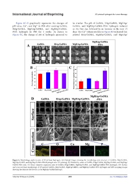
Figure 6. Morphology and structure of 3D-printed hydrogels. (A) Optical images showing the morphology and structure of GelMA, HAp/GelMA,
MgHAp/GelMA, and MgHAp/GelMA-PDA hydrogels after 3D printing. (B) Printability values of GelMA, HAp/GelMA, MgHAp/GelMA, and MgHAp/
GelMA-PDA inks. (C) Struct diameter expansion rates of GelMA, HAp/GelMA, MgHAp/GelMA, and MgHAp/GelMA-PDA hydrogels. (D) Surface
and cross-sectional morphology of 3D-printed GelMA, HAp/GelMA, MgHAp/GelMA, and MgHAp/GelMA-PDA hydrogels and EDS mapping images
showing the element distribution in the MgHAp/GelMA hydrogel.
Volume 10 Issue 5 (2024) 245 doi: 10.36922/ijb.3526