
Page 291 - IJB-10-5
P. 291
International Journal of Bioprinting 3D printing of collagen II-scaffolds
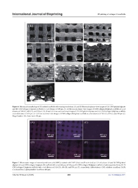
Figure 6. Microscale morphology of 3D-printed scaffolds with varying resolutions. (A and B) Microscale planar-view images of (A) CNF/gelatin/alginate
and (B) CNF/collagen I/alginate scaffolds at a rod distance of 450 μm. (C–E) Microscale planar-view images of CNF/collagen II/alginate scaffolds at a rod
distance of 320 (C), 450 (D), and 550 μm (E). (F and G) Cross-sectional view images of (F) CNF/gelatin/alginate and (G) CNF/collagen I/alginate scaffolds
at a rod distance of 450 μm. (H–J) Cross-sectional view images of CNF/collagen II/alginate scaffolds at a rod distance of 320 (H), 450 (I), and 550 μm (J).
Magnification: 50×. Scale bars: 200 µm.
Figure 7. Microscopic images of mesenchymal stem cells (MSCs) stained with DAPI (blue) and F-actin (red) at 12 h of culture: (A and B) CNF/gelatin/
alginate (A) and CNF/collagen I/alginate (B) scaffolds with a rod distance of 450 μm; (C) CNF/collagen II/alginate scaffold in bulk nonporous form; (D–F)
CNF/collagen II/alginate scaffolds with a rod distance of 320 (D), 450 (E), and 550 μm (F), respectively. Abbreviations: CNF, cellulose nanofiber; DAPI,
4´,6-diamidino-2-phenylindole. Scale bars: 400 µm.
Volume 10 Issue 5 (2024) 283 doi: 10.36922/ijb.3371