
Page 294 - IJB-10-5
P. 294
International Journal of Bioprinting 3D printing of collagen II-scaffolds
At the beginning of cell culture (12 h), cell distribution
was relatively uniform in all scaffolds. However, apparent
cell aggregates (~100 µm) were observed in all collagen II-
based scaffolds on day 4 (96 h), and they were distributed
over the majority of the scaffold area. In contrast, the
aggregates were not observable in gelatin- and collagen
I-based scaffolds. To our knowledge, this work is among
the earliest to successfully visualize the enhancement of
MSC condensation with collagen II.
Quantitatively, the expression of N-cadherin was
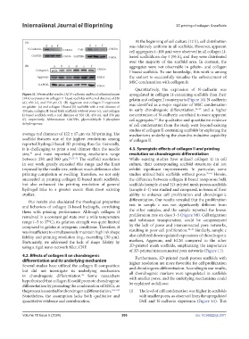
Figure 11. Western blot results. (A) N-cadherin and focal adhesion kinase upregulated in collagen II-containing scaffolds than their
(FAK) expression in collagen II-based scaffolds with a rod distance of 320 gelatin and collagen I counterparts (Figure 10). N-cadherin
(d), 450 (e), and 550 μm (f). (B) Aggrecan and collagen II expression was identified as a major regulator of MSC condensation
on gelatin- (a) and collagen I-based (b) scaffolds with a rod distance of 35–37
450 μm, collagen II-based bulk scaffolds without pores (c), and collagen in early chondrogenic differentiation, and a higher
II-based scaffolds with a rod distance of 320 (d), 450 (e), and 550 μm concentration of N-cadherin correlated to more apparent
(f), respectively. Abbreviation: GADPH, glyceraldehyde 3-phosphate cell aggregates. The qualitative and quantitative evidence
38
dehydrogenase. of cell condensation from the study went beyond existing
studies of collagen II-containing scaffolds by exploring the
average rod diameter of 122 ± 17 μm via 3D printing. The mechanisms underlying the chondro-inductive capability
scaffold features one of the highest resolutions among of collagen II.
reported hydrogel-based 3D printing thus far. Generally,
it is challenging to print a rod thinner than the needle 4.3. Synergistic effects of collagen II and printing
size, and most reported printing resolutions range resolution on chondrogenic differentiation
30
between 250 and 500 μm. 25,31–33 The scaffold resolution While existing studies have utilized collagen II in cell
in our work greatly exceeded this range and the limit culture, their corresponding scaffold structures did not
imposed by the needle size, without much difference after exhibit significant improvements. In particular, some
printing completion or swelling. Therefore, we not only studies utilized bulk scaffolds without pores. 39–41 Herein,
succeeded in printing collagen II-based mesh scaffolds, the difference between collagen II-based nonporous bulk
but also enhanced the printing resolution of general scaffolds (sample c) and 3D-printed mesh porous scaffolds
hydrogel inks to a greater extent than most existing (sample d–f) was studied and compared, in terms of their
studies. ability to enhance cell proliferation and chondrogenic
Our results also elucidated the rheological properties differentiation. Our results revealed that the proliferation
and behaviors of collagen II-based hydrogels, correlating rate in sample c was not significantly different from
them with printing performance. Although collagen II the other samples, and the sample reported the lowest
remained in a constant gel state over a wide temperature proliferation rate on days 3–5 (Figure 9B). Cell migration
range (−5 to 37°C), its gelation strength was quite limited and substance transportation could be compromised
compared to gelatin at cryogenic conditions. Therefore, it by the lack of pores and interconnected pore networks,
42–44
was insufficient to simultaneously maintain high ink shape resulting in poor cell proliferation. Similarly, sample c
fidelity and printing resolution (e.g., exceeding 150 μm). also exhibited downregulated expressions of chondrogenic
Fortunately, we addressed the lack of shape fidelity by markers, Aggrecan, and ECM compared to the other
using a rigid nano-network filler, CNF. 3D-printed mesh scaffolds, emphasizing the importance
of 3D-printed interconnected pore networks (Figure 11).
4.2. Effects of collagen II on chondrogenic Furthermore, 3D-printed mesh porous scaffolds with
differentiation and its underlying mechanism higher resolution are more favorable for cell proliferation
Several studies have utilized the collagen II composition and chondrogenic differentiation. According to our results,
but did not investigate its underlying mechanism all chondrogenic markers were upregulated in scaffolds
in chondrogenic differentiation. Some researchers with smaller pores, and the underlying mechanisms could
22
hypothesized that collagen II could promote chondrogenic be explained as follows:
differentiation by promoting the condensation of MSCs, as
the process is essential for chondrogenic differentiation. 16,21,34 (i) The level of cell condensation was higher in scaffolds
Nonetheless, the assumption lacks both qualitative and with smaller pores, as observed from the upregulated
quantitative evidence and corroboration. FAK and N-cadherin expression (Figure 11). This
Volume 10 Issue 5 (2024) 286 doi: 10.36922/ijb.3371