
Page 293 - IJB-10-5
P. 293
International Journal of Bioprinting 3D printing of collagen II-scaffolds
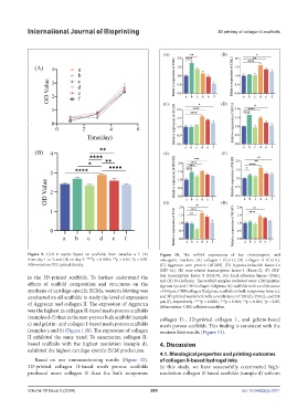
Figure 9. CCK-8 results based on scaffolds from samples a–f: (A) Figure 10. The mRNA expressions of key chondrogenic and
from day 1 to 5; and (B) on day 5. ****p < 0.0001; **p < 0.01; *p < 0.05. osteogenic markers: (A) collagen I (Col-I), (B) collagen II (Col-II),
Abbreviation: OD, optical density. (C) Aggrecan core protein (ACAN), (D) hypoxia-inducible factor-1α
(HIF-1α), (E) runt-related transcription factor-2 (Runx-2), (F) SRY-
in the 3D-printed scaffolds. To further understand the box transcription factor 9 (SOX-9), (G) focal adhesion kinase (FAK),
and (H) N-Cadherin. The scaffold samples evaluated were: CNF/gelatin/
effects of scaffold composition and structures on the alginate (a) and CNF/collagen I/alginate (b) scaffolds with a rod distance
synthesis of cartilage-specific ECMs, western blotting was of 450 μm, CNF/collagen II/alginate scaffolds in bulk nonporous form (c),
conducted on all scaffolds to study the level of expression and 3D-printed mesh form with a rod distance of 320 (d), 450 (e), and 550
of Aggrecan and collagen II. The expression of Aggrecan μm (f), respectively. ****p < 0.0001; ***p < 0.001; **p < 0.001; *p < 0.05.
Abbreviation: CNF, cellulose nanofiber.
was the highest in collagen II-based mesh porous scaffolds
(samples d–f) than in the non-porous bulk scaffold (sample collagen II-, 3D-printed collagen I-, and gelatin-based
c) and gelatin- and collagen I-based mesh porous scaffolds mesh porous scaffolds. This finding is consistent with the
(samples a and b) (Figure 11B). The expression of collagen western blot results (Figure 11).
II exhibited the same trend. To summarize, collagen II-
based scaffolds with the highest resolution (sample d), 4. Discussion
exhibited the highest cartilage-specific ECM production.
4.1. Rheological properties and printing outcomes
Based on our immunostaining results (Figure 12), of collagen II-based hydrogel inks
3D-printed collagen II-based mesh porous scaffolds In this study, we have successfully constructed high-
produced more collagen II than the bulk nonporous resolution collagen II-based scaffolds (sample d) with an
Volume 10 Issue 5 (2024) 285 doi: 10.36922/ijb.3371