
Page 36 - IJB-6-1
P. 36
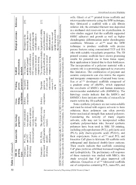
Solvent-based extrusion 3D printing
                                                               cells. Ghosh et al.  printed tissue scaffolds and
                                                                                 [33]
                Reference  [36]       [13]                     microvascular networks using the DIW technique;
                                                               they  fabricated  a  scaffold  with  a  silk  fibroin
                                                      HAVIC: Human aortic valve interstitial cells, PLA: Polylactic acid, MSCs: Mesenchymal stem cells, PCL: Polycaprolactone, HA: Hydroxyapatite, CNT: Carbon nanotubes,
                                                               solution ink; the extruded filament was deposited
                                                               in a methanol-rich reservoir for crystallization. In
                Biological outcomes  HA improved the   bioactivity, there was   good cell adhesion   and spreading at the   scaffold surface in   vitro. The compressive modulus Supported cell viability   and proliferation and   induced osteogenic   differentiation of   hMSCs in vitro and   rapidly integrated with   the tissue in vivo.  vitro studies suggest that the scaffolds supported
                                                               hMSC adhesion  and  growth  as well  as higher
                                                               chondrogenic differentiation under chondrogenic
                                                               conditions.  Miranda  et  al.   used  the  DIW
                                                                                           [34]
                                                               technique  to  produce  scaffolds  with  precise
                                                               porous features using concentrated TCP and HA
                                                               inks with suitable viscoelastic properties. The 3D
                                                               printed ceramic scaffolds have shown promising


                                                               their application is limited due to their brittleness.
                                                               The incorporation  of a polymer  material  with a
                Scaffold   characterization   methods  The compressive   modulus of printed   scaffolds with different   CNT concentrations was   evaluated by uniaxial   compression testing.  of printed scaffolds was   evaluated by uniaxial   compression testing.  results  for potential  use in  bone  tissue  repair;
                                                               ceramic ink is a promising approach to overcome
                                                               this limitation. The combination of polymer and
                                                               ceramic components can also mimic the organic
                                                               and inorganic components of natural bone tissue.
                                                                         [16]
                Printing   process   parameters  No specific   mentioned  The print   speed was   15 cm/s, and   extrusion rates   were as 275   cm 3 /h  Sun  et al.   developed  scaffolds  composed  of
                                                               a  gradient  array  of silk/HA, which  supported
                                                               the cocultures of hMSCs  and human mammary
                                                               microvascular  endothelial  cells  (hMMECs). The
                                                               histology results indicate  that the hMSCs  and


                Cross-  linking   mechanisms  Solvent   evaporation  Solvent   evaporation  hMMECs form intricate networks of extracellular
                                                               matrix within the 3D scaffolds.
                                                                 Some synthetic polymers are not water-soluble
                                                               and must be mixed with organic solvents to form
                                                               solutions;  these polymers can  often provide

                Ink rheology   properties  CNT concentration   was adjusted to   achieve an optimum   viscosity between 2.5   and 7 Pa.s.  The optimal viscosity   of 30–35 Pa·s was   reached. PLGA: Poly (lactic-glycolic acid), SBE: Solvent-based extrusion, TE: Tissue engineering, hMSCs: Human mesenchymal stem cells  better mechanical strength than natural polymers.
                                                               Considering the toxicity of many organic
                                                               solvents, cells may not be incorporated  within
                                                               synthetic  polymer-laden inks. Several synthetic
                                                               polymers  have been  used in SBE 3D printing,

                Ink materials  PCL, HA,   and CNT   dissolved in   dichloromethane  PCL, PLGA,   and HA particles   mixed in   dichloromethane.  including polycaprolactone (PCL), polylactic acid
                                                               (PLA), poly  (lactic-glycolic  acid)  (PLGA), and
                                                               their  copolymers.  Serra  et  al.  used PCL and
                                                                                             [35]
                                                               bioactive CaP glass to fabricate 3D scaffolds with
                                                               orthogonal  and  displaced  double-layer  patterns.
            Table 1. (Continued)  SBE 3D   printing   types  Pneumatic-  driven based   3D printing  Pneumatic-  driven based   3D printing  CaP glass particles exhibited increased roughness
                                                               Their  results  indicate  that  scaffolds  containing

                                                               and hydrophilicity. The preliminary cell response
                                                               of these materials was studied with MSCs; this
                                                               study revealed  that CaP glass improved cell
                                                               adhesion. Gonçalves et al.  fabricated scaffolds
                                                                                        [36]
                                                               out of composites containing PCL, nano-HA, and

           32                          International Journal of Bioprinting (2020)–Volume 6, Issue 1