
Page 91 - IJB-6-1
P. 91
Shuai, et al.
A B C D
E F
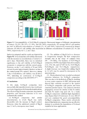
Figure 9. Cytocompatibility of Fe/0.9Mg Si composite: fluorescent images of different concentrations
2
of extract: (A) 0%, (B) 25%, (C) 50%, and (D) 100%, respectively, (E) average MG-63 cell number
per mm in different concentrations of extracts (25, 50, and 100%, respectively) measured by ImageJ
2
software; (F) MG-63 cell viability after incubation in different concentrations of extracts (25, 50, and
100%, respectively) for 1, 2, and 3 days.
further investigated, and the results are depicted in (2) The addition of Mg Si led to a decrease
2
Figure 9F. It was found that cell viability decreased in compression strength, but their compression
with the increase in concentrations of extract at 1 strength was in the range of compact bone
and 2 days. Meanwhile, there was no statistical (90 – 170 MPa). The hardness of Fe/0.9Mg Si
2
significance in the cell viability of Fe/0.9Mg Si composite (145 HV) was higher than other samples
2
composite in comparison with the control group. (112, 134, 142, and 127 HV for Fe, Fe/0.3Mg Si,
2
However, on day 3, cell viability of 50 and 100% Fe/0.6Mg Si, and Fe/1.2Mg Si, respectively).
2
2
extracts became approximately identical as that of A slight decrease was found after adding Mg Si
2
the control group (0% extract). Moreover, during to 1.2 wt% may be caused by micropores in the
3 days of incubation, cell viability was all above microstructure.
70%, indicating no cytotoxicity of Fe/Mg Si (3) Electrochemical tests revealed accelerated
2
composites according to the ISO10993-5 . corrosion behaviors for Fe/Mg Si composites.
[51]
2
This was due to the hydrolysis of Mg Si, resulting
2
4 Conclusions in more matrix exposure to SBF. Immersion tests
showed that corrosion occurred in the interior
In this study, Fe/Mg Si composites were of Fe/Mg Si composites with porous and loose
2
2
successfully fabricated by selective laser melting to corrosion product layers. This induced corrosion
accelerate degradation for biomedical applications. propagation toward the interior of the Fe matrix
The effects of Mg Si on microstructure, mechanical and bulk Fe matrix may peel off. As a result,
2
properties, and corrosion behaviors of Fe were Fe/Mg Si composites exhibited high degradation
2
systematically studied. rates (higher than 0.30 mm/year). In vitro, MG-63
(1) Fe/Mg Si composites had similar cell tests confirmed the good cytocompatibility
2
microstructure, in which Mg Si was easily Fe/Mg Si composites. These results collectively
2
2
discernible with the increase in Mg Si content. showed that hydrolytic expansion was an effective
2
However, when the additional content of Mg Si strategy to accelerate the degradation of Fe-based
2
was 1.2 wt.%, micropores were found. implants for bone applications.
International Journal of Bioprinting (2020)–Volume 6, Issue 1 87