
Page 95 - IJB-6-2
P. 95
Kolan, et al.
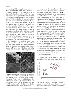
corresponding higher magnification images are in 1 week) irrespective of architecture. This was
shown in Figures 5B and D. These images show due to the higher surface area per unit volume and
the rounded spheroid-like formations on the borate large pores, all measuring about ~1 mm (Table 1),
glass scaffold surface, unlike needle-like crystal that increased the efficiency of the ionic exchange
formations on silicate glass scaffold surface, similar with SBF and made the degradation process more
to observations made by other researchers with these rapid. To comprehend the trends in scaffolds with
glasses [5,35] . The needle-like formations on silicate lower porosities, the unit cell surface area of each
glass surface were confirmed as crystalline HA using architecture (based on the CAD model) was plotted
XRD, whereas spheroid-like formations on borate against the percentage strength reduction, as shown
glass surface were not confirmed to any known in Figure 6. Overall, the plot indicated a higher
crystalline formations. This behavior of borate glass strength reduction with increasing surface area to
is not uncommon as it was reported earlier that it volume ratio (SA/V). The SA/V data points for
could take more than 60 days to form a crystalline gyroid and diamond scaffolds form a distinguishable
calcium phosphate layer in SBF and it also depends group from cubic, spherical, and X scaffolds.
on the strut size [48,49] . This was also observed in However, for a given SA/V ratio (for example, SA/V
some previous work which found the presence of ratio of 2), diamond scaffolds show higher strength
Ca and PO on the reacted surface of the borate reduction in comparison to cubic and spherical
4-
2+
3
glass, indicating an amorphous calcium phosphate scaffolds despite having a similar porosity. This
or carbonate-substituted apatite formation [50,51] . indicates that the lattice structure and pore geometry
The degradation of scaffolds made with do play an important role in controlling scaffold
resorbable materials depends on the material degradation, especially in the case of scaffolds
composition and porosity. Scaffold degradation is made with bioresorbable materials such as bioactive
related to its surface area and the type of soaking glasses. Moreover, it should be noted that the actual
solution (SBF in this study). Scaffolds with higher SA/V values would be higher than the CAD values
porosity degraded the most (~90% strength reduction because of the surface roughness that is inherent to
parts fabricated using the SLS process.
A B
3.4 New bone formation
Scaffolds were firmly integrated with the
surrounding bone and pores were filled with
C D
Figure 5. Scanning electron microscopy images of
borate glass and silicate glass scaffolds at low and
high magnifications after immersion in simulated
body fluids for 1 week: (A and B) Borate glass
outer surface morphology and rounded, irregular
spheroid-like formations, (C and D) silicate glass Figure 6. Percentage reduction in compressive
scaffold surface and well defined needle-like strength of a scaffold versus the ratio of total
crystalline structures at higher magnification. surface area to volume of different architectures.
International Journal of Bioprinting (2020)–Volume 6, Issue 2 91