
Page 98 - IJB-6-2
P. 98
3D-printed borate glass scaffolds for bone repair
soft tissue [52,53] . As expected, addition of BMP-2 3D-printed scaffolds and reported approximately
significantly increased new bone formation to 6% new bone formation . The material extrusion
[55]
almost 40%, based on the total defect area, and 3D-printed scaffolds had pores in the range 150 –
almost filled the entirety of the pores. Our results 300 µm unlike the 1 mm pores of the cubic and
are consistent with a recent study that showed the diamond scaffolds used in this study. Other studies
addition of BMP-2 at the same concentration (1 µg mentioned in Table 4 utilized scaffolds made with
per defect) to defects treated with HA microspheres polymer foam replication technique, freeze-drying
for the same time frame also created approximately technique, and pressed short fiber technique that
40% new bone in a 4.6 mm diameter rat calvarial have significantly smaller pore sizes, thinner strut
defect . The above study investigated the role diameters, and higher porosities. The amount
[54]
of relaxin, a pregnancy hormone, to control and of bone formation in terms of defect region was
enhance BMP-2 release to reduce the need for reported in the range of 9 – 28% after 12 weeks
high concentrations of BMP-2. It was reported of implantation. A relatively higher amount of
that the addition of 0.05 µg of relaxin to 0.5 µg of bone formation (up to 30%) was reported when
BMP-2 induced higher bone formation compared scaffolds were made with copper and zinc doped
to 1 µg of BMP-2 alone per defect. In the future, a borate glass. Higher bone growth for all the above
combination of relaxin and BMP-2 together with scaffolds could be due to smaller pore size range
biomimetic architecture (such as diamond) could and higher porosities compared to the scaffolds
further enhance bone regeneration and repair. used in this study, which were limited by the laser
As new bone formation was about 6% for spot size of the SLS process. The role of pore size
both scaffold types without BMP-2, the results is not completely understood as there exist reports
obtained in this study are largely in agreement with mixed results when using scaffolds with a
with in vivo assessments using borate-based range of pore sizes for bone regeneration . While
[28]
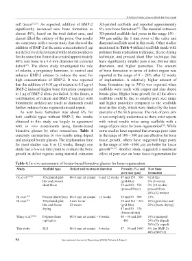
bioactive glasses by other researchers. Table 4 some studies have reported that average pore sizes
concisely summarizes in vivo results using doped in the range of 100 – 300 µm are effective for bone
and undoped borate glasses. The implantation time tissue growth, others have suggested large pores
for most studies was 8 or 12 weeks, though one in the range of 600 –1000 µm are better for tissue
study had a 6-week time point to evaluate the bone growth [56,57] . Another study suggested a nonlinear
growth in defect regions using material extrusion effect of pore size on bone tissue regeneration for
Table 4. In vivo assessment of borate-based bioactive glasses for bone regeneration.
Study Scaffold type Defect and treatment duration Porosity (%) and New bone
pore size (µm) formation
Gu et al. [55,58] 3D-printed grid- Φ4.6 mm rat cranial – 6 and 12 weeks 47 and 150 – 300 Grid-like
like and pressed (grid-like) 6% (6 weeks)
short fibers 58 and 50 – 550 9% (12 weeks)
(pressed fibers) pressed fibers
20% (12 weeks)
Bi et al. [30] Pressed short fibers Φ4.6 mm rat cranial – 12 weeks 58 and 50 – 500 15%
Bi et al. [59] 3D-printed grid- 6 mm femur 50 and 140 – 250 26% (grid-like) and
like and freeze 12 weeks (grid-like) 28% (freeze drying)
drying 47 and 50 – 150
(freeze drying)
Wang et al. [60,61] Polymer foam Φ5.0 mm rat cranial – 8 weeks 80 – 89 and 200 16% (undoped)
replication – 400 29% (Zn doped)
30% (Cu coped)
This study SLS Φ4.6 mm rat cranial – 6 weeks 47 – 54 and 1000 6% (no BMP-2)
40% (BMP-2)
94 International Journal of Bioprinting (2020)–Volume 6, Issue 2