
Page 96 - IJB-6-2
P. 96
3D-printed borate glass scaffolds for bone repair
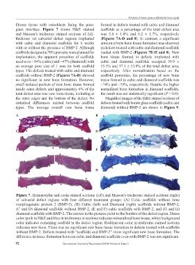
fibrous tissue with osteoblasts lining the pore- formed in defects treated with cubic and diamond
glass interface. Figure 7 shows H&E stained scaffolds as a percentage of the total defect area
and Masson’s trichrome stained sections of full- was 5.8 ± 1.0% and 6.2 ± 1.7%, respectively
thickness rat calvarial defect regions implanted (Figures 7A-D and 8). In contrast, a significant
with cubic and diamond scaffolds for 6 weeks amount of new bone tissue formation was observed
with or without the presence of BMP-2. Although in defects treated with cubic and diamond scaffolds
scaffolds designed at 70% porosity were planned for loaded with BMP-2 (Figures 7E-H and 8). New
implantation, the apparent porosities of scaffolds bone tissue formed in defects implanted with
used were ~54% (cubic) and ~47% (diamond) with cubic and diamond scaffolds occupied 39.9 ±
an average pore size of 1 mm for both scaffold 15.1% and 37.1 ± 13.8% of the total defect area,
types. The defects treated with cubic and diamond respectively. After normalization based on the
scaffolds without BMP-2 (Figures 7A-D) showed scaffold porosities, the percentage of new bone
no significant in new bone formation. However, tissue formed in cubic and diamond scaffolds was
small isolated pockets of new bone tissue formed ~74% and ~79%, respectively. Despite the higher
inside some defects and approximately 6% of the normalized bone formation in diamond scaffolds,
total defect area was new bone tissue, including at the result was not statistically significant (P = 0.8).
the outer edges and the bottom of the defect. No Magnified images of the H&E stained sections of
statistical differences existed between scaffold defects treated with borate glass scaffolds (cubic and
types. The average overall new bone tissue diamond) without BMP-2 are shown in Figure 9.
A B
C D
E F
G H
Figure 7. Hematoxylin and eosin stained sections (left) and Masson’s trichrome stained sections (right)
of calvarial defect regions with four different treatment groups: (A) Cubic scaffolds without bone
morphogenetic protein 2 (BMP-2), (B) Cubic (left) and Diamond (right) scaffolds without BMP-2,
(C and D) diamond scaffolds without BMP-2, (E and F) cubic scaffolds with BMP-2, and (G and H)
diamond scaffolds with BMP-2. The arrows in the pictures point to the borders of the defect region. Dense
color (pink in H&E and blue in trichrome) in sections indicates mineralized bone tissue, white/background
color indicates remaining scaffold in the defect region. Red/maroon color in trichrome stained sections
indicates new bone. There was no significant new bone tissue formation in defects treated with scaffolds
without BMP-2. Defects treated with “scaffolds and BMP-2” show significant new bone formation. The
difference in tissue formation between cubic and diamond scaffolds even with BMP-2 was not significant.
92 International Journal of Bioprinting (2020)–Volume 6, Issue 2