
Page 117 - IJB-6-4
P. 117
Matsugaki, et al.
A
B
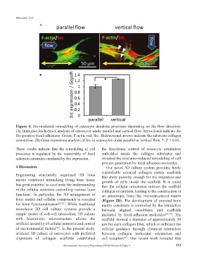
Figure 4. Src-mediated remodeling of osteocyte dendritic processes depending on the flow direction.
(A) Immunocytochemical analysis of osteocytes under parallel and vertical flow. Arrowheads indicate the
Src-positive focal adhesions. Green; F-actin, red; Src. Bidirectional arrows indicate the substrate collagen
orientation. (B) Gene expression analysis of Src in osteocytes under parallel or vertical flow. *: P < 0.05.
These results indicate that the remodeling of cell the directional control of osteocyte orientation
processes is regulated by the reassembly of focal embedded inside the collagen substrates and
adhesion constructs mediated by Src expression. revealed the mechano-induced remodeling of cell
process generation by focal adhesion molecules.
4 Discussion Our novel 3D culture system provides freely
controllable oriented collagen matrix scaffolds
Engineering structurally organized 3D bone that show porosity enough for the migration and
matrix constructs mimicking living bone tissue growth of cells inside the scaffold. It is noted
has great potential to accelerate the understanding that the cellular orientation mirrors the scaffold
of the cellular activities controlling various bone collagen orientation, leading to the construction of
functions. In particular, the 3D arrangement of an anisotropic bone-like microstructured matrix
bone matrix and cellular components is essential (Figure 2D). The development of oriented bone
for bone functionalization [24,25] . While traditional matrix constructs is controlled by the interaction
monolayer 2D cell culture systems provide a between aligned osteoblasts and scaffolds
simple model of cell-cell interaction, 3D culture mediated by focal adhesion molecules [18,19] . The
with biomimetic microstructure allows the scaffold showed a diameter of approximately 50
artificial assembly of cellular networks and control µm for each collagen fiber, which is sufficient for
of environmental factors . In the present study, cellular guidance through chemical interaction
[26]
artificial 3D culture of osteocytes with preferred between collagen molecular orientation and
alignment of collagen scaffolds established cell receptors . Our recent work revealed that
[27]
International Journal of Bioprinting (2020)–Volume 6, Issue 4 113