
Page 18 - IJB-6-4
P. 18
Bioprinting, microfluidics, and organoids to defeat COVID-19
industrial production of diagnostic reagents, they are native tissues with relevant morphology.
vaccines, etc., due to a simple scaling procedure However, their application is significantly limited
[40]
V. However, it was significantly improved by because of low availability and shortage of donor
adding microcarriers – small particles of a cell materials, short viability, and rapid necrosis .
[50]
adhesive substrate (e.g., Cytodex 3). Such method Scaffold- and hydrogel-based models can
modification was approved for the production provide a 3D microenvironment that mimics
of a RSV vaccine and research on virus-host conditions in vivo for cells. Biomaterials that
[43]
interactions . ensure necessary cell-matrix interactions and
[44]
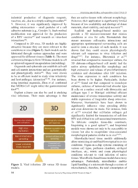
Compared to 2D ones, 3D models are highly appropriate spatiotemporal surrounding cells are
attractive because they are more relevant to the used to form a structure of such models. It was
conditions in vivo (Figure 2). Such models can be shown that they could ensure physiologically
fabricated through various approaches and were relevant cell responses to virus infection and
approved for different viruses (Table 1). The most drugs [39,51] . For instance, Bhowmick et al.
[39]
common technique to form 3D tissue models is cell revealed that compared to monolayer culture, the
or spheroid/organoid encapsulation (embedding). 3D chitosan-collagen-based cell model had the
Organoids and spheroids can establish cell-cell native airway epithelium-like morphology and
and cell-matrix interactions and are genotypically high expression and release of pro-inflammatory
and phenotypically stable . They were shown cytokines and chemokines after IAV infection.
[45]
to be an efficient model to study virus infectivity The virus expression in such conditions has
and host-pathogen interaction [46-48] . For instance, been shown to be higher. Particularly, Archer
using intestinal organoids, Zhou et al. confirmed et al. found out that compared to monolayer
[42]
that MERS-CoV might infect the gastrointestinal cultures, cultures of tumor-derived alveolar type
tract . II cells on a surface coated with fibronectin and
[49]
Explant cultures can also be used in studying collagen type I or Matrigel exhibited efficient
viral infections. Their main advantage is that maintenance of reverse transcriptase activity and
stable expression of Jaagsiekte sheep retrovirus.
Moreover, biomaterials have been shown to
significantly influence virus spreading ability
and even determine its mode. For instance, Imle
et al. revealed that cell-laden collagen gel
[52]
significantly limited the transmission of cell-free
HIV and shifted it to cell-associated transmission.
To fabricate complex tissue-like constructs,
bioprinting is a good option , and bioprinted
[53]
models were shown not only to be susceptible to
viruses but also to recapitulate virus-associated
morphological patterns similar to in vivo [54,55] .
Microfluidic-based tissue models additionally
allow mimicking air and fluid flows typical to in vivo
conditions. Organ-on-a-chip systems consisting of
various cell types, perfusion chambers, air-liquid
interfaces, etc., mimic and create physiological
conditions relevant to viral infection of native
tissues. Microfluidic-based tissue models have many
advantages. Particularly, microfluidics enables
Figure 2. Viral infection: 2D versus 3D tissue liquid handling at a microscale through a system of
models. microchannels; therefore, the total consumption of
14 International Journal of Bioprinting (2020)–Volume 6, Issue 4