Page 386 - IJB-10-6
P. 386
International Journal of Bioprinting                         Innovative manufacturing of ω-3-enriched chocolate


            compression testing machine (EZ-LX; Shimadzu, Japan),   spectrophotometer 6 min after initial mixing. To conduct
            the mechanical strength of the 3D-printed chocolates   the CUPRAC assay, 1 mL portions of CuCl (0.01 M),
                                                                                                    2
            was determined.                                    ammonium acetate buffer (1 M, pH 7.0), and neocuproine
                                                               (7.5 mM) were mixed. After adding 0.1 mL of the extract and
            2.10. Differential scanning calorimetry            1 mL of distilled water, the mixture was incubated at room
            Differential scanning calorimetry (DSC) was performed   temperature for 1 h in the dark. Absorbance measurements
            using DSC-6O Plus (Shimadzu, Japan) to examine     were obtained at 450 nm using a spectrophotometer. The
            the thermal properties of the chocolates (as described   results are expressed as μmol Trolox equivalent (TE)/g dw.
            by  Aydin  et  al. );  a  heating  rate  of  10°C/min  over  a
                         29
            temperature range of 25–350°C in an N  stream was used   2.13. In vitro simulation of gastrointestinal digestion
                                            2
            in the study. The parameters representing the melting   In vitro  simulated  digestion  studies  were  performed  as
            profile associated with the samples, i.e., onset temperature   described by Tolve et al.  and Ozkan et al.  Briefly, 1 g
                                                                                   31
                                                                                                   32
            (T onset   ; the  temperature  at  which  the  samples  begin  to   of chocolate samples was mixed with 0.7 mL of simulated
            melt), peak temperature (T peak ; the temperature at which   salivary fluid (SSF), 5 µL of 3 M CaCl , 195 µL of water,
                                                                                              2
            the highest melting rate was noted), end temperature    and 0.1 mL of α-amylase. After 3 min, 1.5 mL of simulated
            (T ; the temperature at which the samples completely   gastric fluid (SGF), 139 µL of water, 1 µL of 0.3 M CaCl ,
              end
                                                                                                            2
            melted), and ΔH (the energy that is required for complete   and 0.32 mL of pepsin were added. The pH of the solution
            sample melting) were determined from thermograms   was subsequently adjusted to 3.0 with 40 µL of HCl (1
            obtained from the measurements.                    M); the sample was incubated at 37°C for 2 h. After 2 h
            2.11. Determination of total polyphenol content    of incubation at 37°C, 1.3 mL of simulated intestinal fluid
            The chocolate samples were defatted prior to phenolic   (SIF), 1 mL of lipase, 1 mL of pancreatin, 0.5 mL of bile
            extraction using the approach described by Hu et al.    salts, 8 µL of 0.3 M CaCl , and 162 µL of water were added.
                                                         30
                                                                                  2
            with some modifications. Briefly, the samples were sliced   The pH was adjusted to a pH value of 7 in this phase using
            into small pieces; 0.5 g of each piece was extracted using   30 µL of NaOH (1 M). The sample was incubated at 37°C for
            2.5 mL of n-hexane twice via shaking for 45 min at 25 ±   3 h. The aliquots collected at the end of the salivary, gastric,
            0.03°C at 150 ×g. After each extraction step, the mixtures   and intestinal phases were added to 9 mL of methanol/
            were centrifuged at 2000 ×g for 10 min; the supernatant   HCl/water (79.9:0.1:20, v/v/v) and brought to −20°C in
            was subsequently discarded. After the second extraction   order to inhibit the enzyme activity prior to polyphenol
            step, the samples were dried in a hot-air dryer for 24 h at   determination. A blank test tube without a chocolate
            25°C.  The defatted samples  were  subsequently  weighed;   sample but with all digestion fluids was also analyzed. The
            the phenolics were extracted using 5 mL of 0.1% HCl   bioaccessibility index (BI) value was determined as follows:
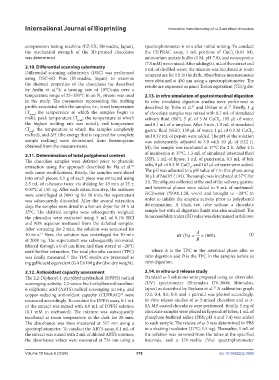
            and 80% aqueous methanol from the defatted samples.
            After vortexing for 2 min, the solution was sonicated for
            20 min.  Next, the solution was centrifuged for 10 min          BI (%) =  A  × 100 %           (I)
                  31
            at 2000 ×g. The supernatant was subsequently recovered,                  B
            filtered through a 0.45 μm filter, and then stored at −20°C
            until further extraction. The total phenolic content (TPC)   where  A is the TPC in the intestinal phase after  in
            was finally measured.  The TPC results are presented as   vitro digestion and B is the TPC in the samples before in
                             32
            mg gallic acid equivalent (GAE)/100 g dw (dw: dry weight).  vitro digestion.
            2.12. Antioxidant capacity assessment              2.14. In vitro ω-3 release study
            The 2,2-Diphenyl-1-picrylhydrazylradical (DPPH) radical   Standard ω-3 solutions were prepared using an ultraviolet
            scavenging activity, 2,2-azino-bis-3-ethylbenzothiazoline-  (UV)  spectrometer  (Shimadzu  UV-3600; Shimadzu,
                                                                                            33
            6-sulphonic acid (ABTS) radical scavenging activity, and   Japan) as described by Baykara et al.  A calibration graph
            copper-reducing antioxidant capacity (CUPRAC)   were   (0.2, 0.4, 0.6, 0.8, and 1 µL/mL) was plotted accordingly.
                                                    32
            measured accordingly. To conduct the DPPH assay, 0.1 mL   In vitro release studies of ω-3-mixed chocolate and ω-3-
            of the extract was mixed with 4.9 mL of DPPH solution   SA MP-coated chocolates were performed. Briefly, 5 mg of
            (0.1  mM  in  methanol).  The  mixture  was  subsequently   chocolate samples were placed in Eppendorf tubes; 1 mL of
            incubated  at  room  temperature  in  the  dark  for  20  min.   phosphate-buffered saline (PBS; pH 4 and 7.4) was added
            The absorbance was then measured at 517 nm using a   to each sample. The release of ω-3 was determined in PBS
            spectrophotometer. To conduct the ABTS assay, 0.1 mL of   in a shaking incubator (37°C; 1.5 ×g). Thereafter, 1 mL of
            the extract was mixed with 2 mL of diluted ABTS solution;   the solution was removed from the tubes at the specified
            the absorbance values were measured at 734 nm using a   intervals, and a UV-visible (Vis) spectrophotometer

            Volume 10 Issue 6 (2024)                       378                                doi: 10.36922/ijb.3969

