
Page 403 - IJB-10-6
P. 403
International Journal of Bioprinting 3D-printed PCL-MNP multifunctional scaffolds
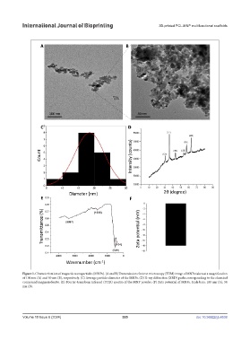
Figure 1. Characterization of magnetic nanoparticles (MNPs). (A and B) Transmission electron microscopy (TEM) image of MNPs taken at a magnification
of 100 nm (A) and 50 nm (B), respectively. (C) Average particle diameter of the MNPs. (D) X-ray diffraction (XRD) peaks corresponding to the chemical
compound magnesioferrite. (E) Fourier-transform infrared (FTIR) spectra of the MNP powder. (F) Zeta potential of MNPs. Scale bars: 100 nm (A); 50
nm (B).
Volume 10 Issue 6 (2024) 395 doi: 10.36922/ijb.4538