Page 404 - IJB-10-6
P. 404
International Journal of Bioprinting                             3D-printed PCL-MNP multifunctional scaffolds


            (Figure  1E). The XRD analysis allowed us to determine   Once the MNPs were characterized, scaffold preparation
            the concentration of different elements in the sample.   was carried out. A 40% (w/v) solution of PCL was used
            Figure 1D displays peaks at 2Ѳ = 31, 36, 43, 54, 57, and   as the base material. To this solution, 0–2.5 g MNPs were
            63. XRD analysis was conducted through the Execute   added to obtain 0–50% (w/v) PCL-MNP solutions (0%,
            Search and Match tool, and a match with magnesioferrite   2.5%, 5%, 10%, 20%, 30%, and 50%). The printing process
            was observed. The FTIR spectra in Figure 1E revealed the   is represented in Figure 2A. It can be observed from the
            chemical bonds in the MNPs. A sharp peak was observed   printed scaffold that the extrusion 3D printing process was
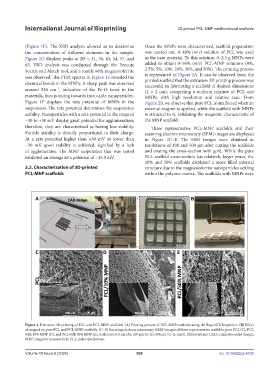
                                                               successful in fabricating a scaffold of desired dimensions
                         −1
            around 540 cm , indicative of the Fe-O bond in the   (2 × 2 cm), comprising a uniform mixture of PCL and
            materials, thus pointing towards iron oxide nanoparticles.   MNPs,  with  high  resolution  and  relative  ease.  From
            Figure  1F displays the zeta potential of MNPs in the   Figure 2B, we observe that pure PCL is unaffected when an
            suspension. The zeta potential determines the suspension   external magnet is applied, while the scaffold with MNPs
            stability. Nanoparticles with a zeta potential in the range of   is attracted to it, validating the magnetic characteristic of
            −30 to +30 mV display great potential for agglomeration;   the MNP scaffold.
            therefore, they are characterized as having low stability.   Three representative PCL-MNP scaffolds and their
            Particle stability is directly proportional to their charge.   scanning electron microscopy (SEM) images are displayed
            At  a zeta potential  higher  than +30  mV  or  lower  than   in  Figure  2C–E.  The  SEM images  were  obtained  at
            −30  mV,  good  stability  is  achieved,  signified  by  a  lack   resolutions of 100 and 500 µm after cutting the scaffolds
            of agglomerates. The MNP  suspension that was  tested   and coating the cross-section with gold. While the pure
            exhibited an average zeta potential of −15.9 mV.    PCL scaffold cross-section has relatively larger pores, the
                                                               10% and 50% scaffolds displayed a more filled internal
            3.2. Characterization of 3D-printed                structure due to the magnesioferrite nanoparticles settling
            PCL-MNP scaffolds                                  within the polymer matrix. The scaffolds with MNPs were


            Figure 2. Extrusion 3D printing of PCL and PCL-MNP scaffolds. (A) Printing process of PCL-MNP scaffolds using the RegenHU bioprinter. (B) Effect
            of magnet on pure PCL and PCL-MNP scaffolds. (C–E) Scanning electron microscopy (SEM) images of three representative scaffolds: pure PCL (C), PCL
            with 10% MNP (D), and PCL with 50% MNP (E). Scale bars: 0.5 cm (A); 100 µm (C–E); 500 µm (C–E, inset). Abbreviations: CAD, computer-aided design;
            MNP, magnetic nanoparticle; PCL, polycaprolactone.


            Volume 10 Issue 6 (2024)                       396                                doi: 10.36922/ijb.4538

