
Page 409 - IJB-10-6
P. 409
International Journal of Bioprinting 3D-printed PCL-MNP multifunctional scaffolds
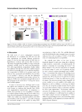
Figure 6. Cell-laden scaffolds in AMF. (A) Schematic of seeding human mesenchymal stem cells (hMSCs) and bone cancer cells (BCCs) in pure
polycaprolactone (PCL) and PCL with 50% magnetic nanoparticles (MNPs) scaffolds placed in an alternating magnetic field (AMF). (B) Alamar Blue
analysis of hMSCs and BCCs after being placed in an AMF. *p < 0.05. Abbreviation: NS, non-significant.
4. Discussion concentrations as high as 50%. The scaffolds fabricated
with 50% MNPs helped achieve strength close to the native
The dual needs of cancer management necessitate
the fabrication of a material that will not only aid trabecular bone, with a modulus of 229.06 ± 37.05 MPa,
bone regeneration and provide adequate mechanical while still maintaining their biocompatibility.
support in vivo but also target tumors and kill them via The critically sized defect of the bone is often
hyperthermia treatment and prevent their recurrence. irregularly shaped. In such cases, being able to fabricate
This study investigates the possibility of using a composite a high-resolution, customizable, and patient-specific 3D
polymeric scaffold, made by combining biocompatible implant is important for overcoming this barrier. The
40
PCL with magnesioferrite nanoparticles. Earlier studies design features of the critically sized defect can typically
have indicated that the PCL and IONP scaffolds support be extracted using CT scans, and 3D printing can then
cell adhesion and proliferation and are thus biocompatible; be used to mimic a patient-customized bone scaffold.
41
the incorporation of IONPs in the polymer matrix also
enhances the mechanical and wettability characteristics Figure 2 demonstrates the extrusion 3D printing process
of the scaffold. 40,41 However, even after the substantial to fabricate a composite scaffold of desired dimensions
improvement in mechanical characteristics achieved by and pore size. While the pores of the grid-like structures
using the IONPs, their mechanical strength falls short of printed were about 5 × 5 mm, they only serve as a proof-
that possessed by the native bone, limiting their use as of-concept. The extrusion process using a RegenHU 3D
30
bone scaffold implants. printer enabled us to print scaffolds with smaller pore
diameters as well. In addition to achieving the desired
In order to overcome these challenges and to make
the scaffold more suitable for cancer management, this pore size, our composite samples revealed that the pores
study focuses on replacing the typically used IONPs were quite interconnected, which is a necessary condition
43
with magnesioferrite nanoparticles. In comparison to for bone tissue regeneration. Furthermore, while the
IONPs, these particles are less susceptible to oxidation extrusion 3D printing process is indeed relatively easy
and display a greater heating capacity, which makes them to use and implement, it is still limited by its inability to
more attractive alternatives for hyperthermia treatment. form highly complex structural configurations. 44,45 Other
42
While other studies only fabricated scaffolds with MNP additive manufacturing processes, such as fused deposition
concentrations of less than 15%, 17,30 this study enhanced modeling (FDM), can be explored in the future with PCL-
the mechanical strength of the scaffold by using MNP MNP composite filaments.
Volume 10 Issue 6 (2024) 401 doi: 10.36922/ijb.4538