
Page 408 - IJB-10-6
P. 408
International Journal of Bioprinting 3D-printed PCL-MNP multifunctional scaffolds
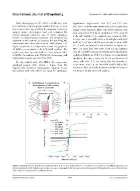
When developing our PCL-MNP scaffolds, we aimed hyperthermia experiments. Pure PCL and PCL with
to (i) fabricate a biocompatible scaffold that aids in bone 50% MNP scaffolds were seeded with hMSCs and bone
tissue regeneration by providing the necessary mechanical cancer cells in respective glass vials. These scaffolds were
support under physiological load and activating the then cultured for 24 h in an incubator at 37°C. After 24
correct signaling pathways, and (ii) target cancerous h, the cell viability of the scaffolds was measured. After
tumors via hyperthermia treatment. The hyperthermia the supernatant was collected in a 96-well plate and fresh
capability of the scaffolds is evaluated by measuring the
temperature rise when placed in an AMF (Figure 5A). media replaced, the scaffolds were then placed in an AMF
Figure 5B presents the temperature versus time graph for for 2 h each, as depicted in the schematic in Figure 6A.
all MNP concentrations in the PCL-MNP scaffolds. The After 2 h, these glass vials were taken out and supplied
peak temperature increases with increasing concentration with AB to identify changes in viability after subjecting the
of MNPs. For scaffolds with 50% MNPs, the average peak seeded scaffolds to an AMF. From Figure 6B, a statistically
temperature reached was more than 46 C. significant decrease is observed in the proliferation of
o
For the scaffold with 50% MNPs, the temperature cancer cells after 2 h, indicating that the increase in
stabilized around 46°C, which is higher than the temperature caused by the 50% MNP scaffold helped kill
hypothermia threshold temperature required; hence, the cancer cells. Conversely, the hMSCs exhibited increased
the scaffold with 50% MNPs was used for subsequent proliferation for the 50% MNP scaffold.
Figure 5. Magnetization and hyperthermic measurements of PCL and PCL-MNP scaffolds. (A) Schematic of the process of a scaffold being placed in an
alternating magnetic field, while the temperature is measured using a probe. (B) Temperature versus time graph. (C) Magnetization curves. Abbreviations:
MNP, magnetic nanoparticle; PCL, polycaprolactone.
Volume 10 Issue 6 (2024) 400 doi: 10.36922/ijb.4538