Page 543 - IJB-10-6
P. 543
International Journal of Bioprinting                              Design and property of PLPG/PDLA scaffold


            Testing Machine Co., LTD, China) at a compression speed   washed with phosphate-buffered saline (PBS) and analyzed
            of 1 mm/min. Each experiment had three parallel samples.  using a fluorescence microscope (LEICA DMi8; LEICA,
                                                               Germany). Each experiment had three parallel samples.
            2.6. Degradation behavior of the
            PLPG/PDLA scaffolds                                2.7.3. Immunofluorescent staining
            The enzymatic degradation behavior of the PLPG/PDLA   MC3T3-E1 cells (3 × 10  cells per mL) co-cultured on the
                                                                                  4
            scaffolds was assessed in a proteinase K solution over 15   PLPG/PDLA scaffolds for 12 and 72 h were fixed with
            days. Initially, the PLPG/PDLA scaffolds were immersed in   paraformaldehyde for 0.5 h and then permeabilized with
            3 mL of proteinase K solution containing tris buffer (0.05   Triton X-100 solution. The cells were co-cultured with an
            M; pH 8.5). To maintain enzyme activity, the proteinase   anti-vinculin antibody for 12 h at 4°C. Cell actin  stress
            K-tris buffer was  replaced  every  two  days.  The  scaffolds   fibers were stained using a rhodamine-phalloidin solution
            were removed at specified time points, washed with plenty   for 0.5 h, and cell nuclei were stained with 10 µg/mL DAPI.
            of water, and then placed in a vacuum drying oven (37°C)   The adhesion morphology of the MC3T3-E1 cells cultured
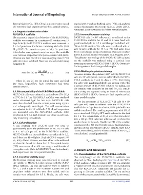
            until their mass stabilized. Mass loss was calculated using   on  the  scaffolds  was  analyzed  using  a  confocal  laser
            Equation II:                                       scanning microscope (LEICA DMi8 S; LEICA, Germany).
                                                               Each experiment had three parallel samples.
                                                               2.7.4. Alkaline phosphatase staining
                   Mass loss (%)           100         (II)
                                                               To assess alkaline phosphatase (ALP) activity, MC3T3-E1
                                                               cells (6 × 10  cells per mL) were co-cultured with the PLPG/
                                                                        4
               where  M  and  M  are the initial dry mass and final   PDLA scaffolds for 7 and 14 days at 37°C. After fixing
                             d
                      i
            dry mass, respectively. Each experiment had three   the cells with paraformaldehyde and rinsing with PBS,
            parallel samples.                                  a certain amount of BCIP/NBT solution was added, and
                                                               the samples were incubated in the dark for 24 h. Finally,
            2.7. Biocompatibility of the PLPG/PDLA scaffolds   the staining was captured using an inverted microscope
            MC3T3-E1 cells were cultured in an incubator (5% CO )   (LEICA DMi8 S; LEICA, Germany). Each experiment had
                                                         2
            at 37°C for 5 h. The PLPG/PDLA scaffolds were sterilized   three parallel samples.
            under ultraviolet light for two days. MC3T3-E1 cells   For the assessment of ALP, MC3T3-E1 cells (6 × 10
                                                                                                             4
            were then detached from the culture plates using trypsin   cells per mL) were co-cultured with the PLPG/PDLA
            and subsequently centrifuged. The cell concentration   scaffolds for 14 days. Post-incubation, RIPA lysis buffer was
            was adjusted to 6 × 10  cells/mL. A 30 µL cell suspension   added to each well. The resulting supernatants with 150 μL
                              5
            was inoculated onto the PLPG/PDLA scaffolds. After   were mixed with 150 μL pNPP and incubated in the dark
            incubation for 6 h, a fresh medium was added to each well,   for 1 h. The supernatants of 20 μL were then introduced
            fully immersing the scaffolds.                     into a 200 μL BCA detection solution and incubated for

            2.7.1. Cell proliferation                          another hour in the dark. Finally, the OD of the solution
            A  cell  counting  kit-8 (CCK-8)  assay  was  used  to   was measured at 405 and 562 nm using a multifunctional
            quantitatively assess the proliferation of MC3T3-E1 cells   enzyme-labeled instrument (TECAN Infinite M200 PRO;
            at 6 × 10  cells per mL in the PLPG/PDLA scaffolds.   Tecan, Switzerland), and the relative activity of ALP (R ALP )
                    4
            MC3T3-E1 cells and the scaffolds were co-cultured for 1, 3,   was calculated using Equation III:
            and 5 days in a 48-well plate. 10 μL of CCK-8 reagent (100
            μL, 10%), diluted 10 times, was then added to the wells and   R ALP  =OD /OD ×100%            (III)
                                                                                 405
                                                                                      562
            incubated in the cell incubator for 2 h. The optical density
            (OD) was measured at 450 nm using a multifunctional
            microplate reader (Infinite M200PRO; Tecan, Switzerland).   3. Results and discussion
            Each experiment had three parallel samples.
                                                               3.1. Characterization of the PLPG/PDLA scaffolds
            2.7.2. Live/dead viability assay                   The morphology of the PLPG/PDLA scaffolds was
            The live/dead staining assay was performed to evaluate   observed via SEM. As displayed in Figure 1a–d, the PLPG/
            the viability of MC3T3-E1 cells cultured with the PLPG/  PDLA scaffolds, with pore sizes of 200–300 μm, exhibited
            PDLA scaffolds. The cells and scaffolds were co-cultured   a regular network structure and uniform struts, indicating
            for 1, 2, and 3 days in the incubator. Calcein-AM and   that the PLPG/PDLA composites are suitable  for 3D
            propidium iodide solutions were then added, and the   printing to fabricate complex scaffolds. The top and side
            samples were cultured for 0.5 h. Finally, the cells were   views of the pore structure inside the scaffold demonstrated

            Volume 10 Issue 6 (2024)                       535                                doi: 10.36922/ijb.4645

