
Page 547 - IJB-10-6
P. 547
International Journal of Bioprinting Design and property of PLPG/PDLA scaffold
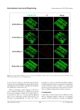
Figure 4. Live/dead staining of MC3T3-E1 cells co-cultured with PLPG/PDLA scaffolds. Scale bars: 50 μm. Abbreviations: PDLA: Poly(D-lactic acid);
PLPG: PLLA-ran-PDO-ran-GA; PI: Propidium iodide.
Z-axis of the pores. Moreover, the density observed in the primarily due to the fact that the PLPG/PDLA scaffolds,
overhead view was much higher than that of the 2D results, being biodegradable polymer materials, have limited ability
indicating that the PLPG/PDLA scaffolds favor the growth to promote bone formation. In light of this, future work
and proliferation of MC3T3-E1 cells. will involve introducing bioactive materials into the PLPG/
PDLA scaffolds to enhance their osteogenic properties.
Alkaline phosphatase (ALP) staining of MC3T3-E1
cells co-cultured on PLPG/PDLA scaffolds was performed 4. Conclusion
to assess early osteogenic activity over 14 days (Figure 6).
As presented in Figure 6a, a few gray deposits appeared In this study, we demonstrated that the addition of PDO
after 14 days. Quantitative analysis indicated that ALP and GA monomers negatively affects the crystallization
expression was approximately 1.8 (Figure 6b). This is capacity of PLPG copolymers, resulting in insufficient
Volume 10 Issue 6 (2024) 539 doi: 10.36922/ijb.4645