
Page 548 - IJB-10-6
P. 548
International Journal of Bioprinting Design and property of PLPG/PDLA scaffold
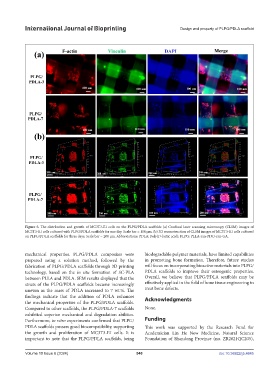
Figure 5. The distribution and growth of MC3T3-E1 cells on the PLPG/PDLA scaffolds (a) Confocal laser scanning microscopy (CLSM) images of
MC3T3-E1 cells cultured with PLPG/PDLA scaffolds for one day. Scale bar = 100 μm. (b) 3D reconstruction of CLSM images of MC3T3-E1 cells cultured
on PLPG/PDLA scaffolds for three days. Scale bar = 200 μm. Abbreviations: PDLA: Poly(D-lactic acid); PLPG: PLLA-ran-PDO-ran-GA.
mechanical properties. PLPG/PDLA composites were biodegradable polymer materials, have limited capabilities
prepared using a solution method, followed by the in promoting bone formation. Therefore, future studies
fabrication of PLPG/PDLA scaffolds through 3D printing will focus on incorporating bioactive materials into PLPG/
technology, based on the in situ formation of SC-PLA PDLA scaffolds to improve their osteogenic properties.
between PLLA and PDLA. SEM results displayed that the Overall, we believe that PLPG/PDLA scaffolds may be
struts of the PLPG/PDLA scaffolds became increasingly effectively applied in the field of bone tissue engineering to
uneven as the mass of PDLA increased to 7 wt.%. The treat bone defects.
findings indicate that the addition of PDLA enhances Acknowledgments
the mechanical properties of the PLPG/PDLA scaffolds.
Compared to other scaffolds, the PLPG/PDLA-7 scaffolds None.
exhibited superior mechanical and degradation abilities.
Furthermore, in vitro experiments confirmed that PLPG/ Funding
PDLA scaffolds possess good biocompatibility, supporting This work was supported by the Research Fund for
the growth and proliferation of MC3T3-E1 cells. It is Academician Lin He New Medicine, Natural Science
important to note that the PLPG/PDLA scaffolds, being Foundation of Shandong Province (no. ZR2021QC205),
Volume 10 Issue 6 (2024) 540 doi: 10.36922/ijb.4645