
Page 546 - IJB-10-6
P. 546
International Journal of Bioprinting Design and property of PLPG/PDLA scaffold
after six days of enzymatic degradation. By day 15, the Furthermore, the viability of MC3T3-E1 cells co-
PLPG/PDLA scaffolds exhibited significant degradation. cultured with PLPG/PDLA scaffolds was assessed using
Additionally, as the PDLA mass increased, numerous voids live/dead staining over five days (Figure 4). Only a few
appeared on the strut surfaces, while the undegraded areas cells stained red (indicating dead cells) were detected,
remained relatively smooth. This can be attributed to the while a large number of living cells stained green attached
fact that proteinase K primarily degraded the amorphous to the struts of the PLPG/PDLA scaffolds, indicating
regions surrounding the spherulites. 35,36 The SEM results that the PLPG/PDLA scaffolds promote the growth and
are consistent with the weight loss data. Therefore, based proliferation of MC3T3-E1 cells.
on the mechanical properties and degradation behavior, The proliferation capacity of MC3T3-E1 cells co-
the PLPG/PDLA-7 scaffolds demonstrate potential for use cultured with PLPG/PDLA scaffolds was further evaluated
in tissue engineering. using confocal laser scanning microscopy (CLSM) images
taken after a day. As displayed in Figure 5a, many cells
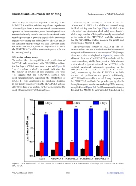
3.3. In vitro cellular assay adhered to the struts of the PLPG/PDLA scaffolds, with the
To analyze the biocompatibility and proliferation of cytoskeleton clearly visible. The expression of the adhesion
MC3T3-E1 cells co-cultured with PLPG/PDLA scaffolds protein vinculin (green) indicated that MC3T3-E1 cells
for five days, a CCK-8 assay was conducted (Figure 3). exhibited substantial cytoplasmic spreading on the
The OD values gradually increased, indicating that PLPG/PDLA scaffolds and were well-attached to F-actin
the number of MC3T3-E1 cells increased over time. (red), demonstrating that the PLPG/PDLA scaffolds
This suggests that the PLPG/PDLA scaffolds have promote cell proliferation and growth. Additionally,
good biocompatibility, supporting the proliferation of MC3T3-E1 cells were able to spread through the pores in
MC3T3-E1 cells. Additionally, no significant difference the PLPG/PDLA scaffolds. The growth capacity of cells
in cell viability was observed in the PLPG/PDLA scaffolds through these pores was also assessed using CLSM scanning
after three days of co-culture, further demonstrating the along the Z-axis (Figure 5b). The 3D reconstruction images
good cytocompatibility of these scaffolds. displayed that MC3T3-E1 cells were distributed along the
Figure 3. CCK-8 assay of MC3T3-E1 cells cultured on PLPG/PDLA scaffolds (n = 3). Abbreviations: PDLA: Poly(D-lactic acid); PLPG: PLLA-ran-
PDO-ran-GA.
Volume 10 Issue 6 (2024) 538 doi: 10.36922/ijb.4645