
Page 110 - IJB-7-1
P. 110
HA15-loaded Bone Tissue Scaffold
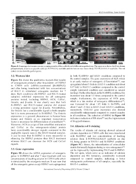
Figure 4. Scanning electron microscopy scanning results of the scaffolds with different magnifications. The upper row shows the β-tricalcium
phosphate/poly (Lactic-co-glycolic acid) (β-TCP/PLGA) scaffolds and the bottom row shows the β-TCP/PLGA/HA15 scaffolds. The red
dotted circles show the perfect pores.
3.2. Western blot in both Si-HSPA5 and HA15 conditions compared to
the control samples. The gene expression of ALP, which
Figure 5A shows the qualitative western blot results is an early marker of osteogenic differentiation , was
[38]
of osteogenesis proteins after knockdown of HSPA5 upregulated about 3 folds in HA15-1 condition and about
in C3H10 cells (siRNA-transfected [Si-HSPA5])
and after being transfected with two concentrations 0.17 fold in HA15-2 condition compared to the control
of HA15 in stimulated osteogenic medium for 7 sample (untreated condition and considered as natural
days. Both conditions (Si-HSPA5- and HA15-treated healing). On the other hand, in the Si-HSPA condition, this
samples) exhibited expression for all osteogenic increment was about 1.5 times compared to the control
proteins tested, including HSPA5, OCN, Col1a1, sample. Furthermore, the expression of OCN gene,
[39]
Osterix, and β-actin. It was clearly seen that both which is a late marker of osteogenic differentiation ,
Si-HSPA5- and HA15-treated samples did express was increased for about 1.85 folds in Si-HSPA, and
a strong persistent signal for β-actin. Nevertheless, about 3 and 1.8 fold in HA15-2 and HA15-1 conditions,
the Osterix has a considerable signal in both samples respectively. COL1A1 gene expression also showed
but it is more pronounced in Si-HSPA5. The Osterix considerable enhancement compared to control samples
expression is a general phenomenon in human bone in all conditions. The reduction of HSPA5 in Figure 5B
[40]
tissues and Osterix as an important transcription indicates a reduction of ER stress and the improvement
factor is necessary for differentiation of osteoblasts [37] . of drug performance.
Moreover, OCN as a sign of late osteoblast maturation 3.4. Alizarin red S staining
and Col1a1 as the osteogenesis-related protein
have considerably stronger signals compared to the The results of alizarin red staining showed enhanced
negligible signals seen in the HA15-treated samples. calcium deposition in C3H10 cells that were transfected
It can be seen that Si-HSPA5 is more successful in with Si-HSPA5 and two concentrations of leaching
silencing the HSPA5 proteins than HA15 treatment solution from β-TCP/PLGA/HA15 scaffolds on day
whereas HA15 can decrease the OCN and Col1a1 14, being greater in HA15-1 and Si-HSPA5 conditions
signals. (Figure 5C). Hence, the mineralization of extracellular
matrix that usually happens during in vitro osteogenesis
[41]
3.3. Gene expression can be improved by implementing HA15 and Si-HSPA5.
Figure 5B shows the mRNA expression of osteogenic The red spots, which serve as an indicator of calcium
genes in Si-HSPA5 and after 7 days treatment with two deposition and HA formation, considerably increased
concentrations of leaching solution in C3H10 cells which compared to control samples, indicating a better
is stimulated by the osteogenic medium. It was seen that osteogenesis performance of all samples, especially the
all the osteogenic genes except HSPA5 were upregulated HA15-1 condition.
106 International Journal of Bioprinting (2021)–Volume 7, Issue 1