
Page 112 - IJB-7-1
P. 112
HA15-loaded Bone Tissue Scaffold
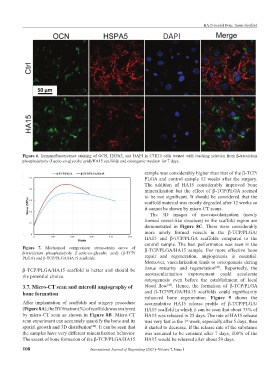
Figure 6. Immunofluorescence staining of OCN, HSPA5, and DAPI in C3H10 cells treated with leaching solution from β-tricalcium
phosphate/poly (Lactic-co-glycolic acid)/HA15 scaffolds and osteogenic medium for 7 days.
sample was considerably higher than that of the β-TCP/
PLGA and control sample 12 weeks after the surgery.
The addition of HA15 considerably improved bone
mineralization but the effect of β-TCP/PLGA seemed
to be not significant. It should be considered that the
scaffold material was mostly degraded after 12 weeks so
it cannot be shown by micro-CT scans.
The 3D images of neovascularization (newly
formed vessel-like structure) in the scaffold region are
demonstrated in Figure 8C. There were considerably
more newly formed vessels in the β-TCP/PLGA/
HA15 and β-TCP/PLGA scaffolds compared to the
control sample. The best performance was seen in the
Figure 7. Mechanical compression stress-strain curve of β-TCP/PLGA/HA15 sample. For more effective bone
β-tricalcium phosphate/poly (Lactic-co-glycolic acid) (β-TCP/
PLGA) and β-TCP/PLGA/HA15 scaffolds. repair and regeneration, angiogenesis is essential.
Moreover, vascularization leads to osteogenesis during
[45]
β-TCP/PLGA/HA15 scaffold is better and should be tissue maturity and regeneration . Reportedly, the
the potential choice. neovascularization improvement could accelerate
osteogenesis even before the establishment of local
3.7. Micro-CT scan and microfil angiography of blood flow . Hence, the formation of β-TCP/PLGA
[46]
bone formation and β-TCP/PLGA/HA15 scaffolds could significantly
enhanced bone regeneration. Figure 9 shows the
After implantation of scaffolds and surgery procedure accumulative HA15 release profile of β-TCP/PLGA/
(Figure 8A), the BV fraction (%) of scaffolds was analyzed HA15 scaffold in which it can be seen that about 73% of
by micro-CT scan as shown in Figure 8B. Micro-CT HA15 was released in 25 days. The rate of HA15 release
scan experiment can accurately quantify the bone and its was very fast in the 1 week, especially after 5 days, then
st
spatial growth and 3D distribution . It can be seen that it started to decrease. If the release rate of the substance
[44]
the samples have very different mineralization behavior. was assumed to be constant after 7 days, 100% of the
The extent of bone formation of the β-TCP/PLGA/HA15 HA15 would be released after about 59 days.
108 International Journal of Bioprinting (2021)–Volume 7, Issue 1