
Page 111 - IJB-7-1
P. 111
Zheng, et al.
A B
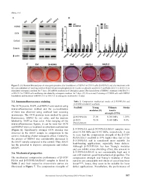
C
Figure 5. (A) Western blot analyses of osteogenic proteins after knockdown of HSPA5 in C3H10 cells (Si-HSPA5) and after treatment with
two concentrations of leaching solution from β-tricalcium phosphate/poly (Lactic-co-glycolic acid)/HA15 scaffolds (HA15-1, HA15-2) in
stimulated osteogenic medium for 7 days. (B) mRNA expression of osteogenic genes after knockdown of HSPA5, treatment with HA15-1
or HA15-2 in C3H10 cells and being stimulated by osteogenic medium for 7 days. (C) Alizarin red S staining of C3H10 cells with HSPA5
knockdown and treatment with HA15-1 or HA15-2 in osteogenic medium for 14 days.
3.5. Immunofluorescence staining Table 2. Compression mechanical results of β-TCP/PLGA and
β-TCP/PLGA/HA15 scaffolds
The OCN protein, DAPI, and HSPA5 were stained using
Young
immunofluorescence method and the co-localization Scaffold modulus (E) Ultimate Strain
tensile
of them was observed using confocal laser scanning strength (UTS)
microscope. The OCN proteins were marked by green
fluorescence, HSPA5 by red color, and the nucleus β-TCP/PLGA 27.86 0.366 MPa 1.28%
labeled by DAPI as blue color. After merging all the β-TCP/ 31.36 0.443 MPa 8.6%
immunofluorescence figures, it can be seen that OCN PLGA/HA15
and HSPA5 were co-localized in cytoplasm and nucleus
(Figure 6). Significantly stronger OCN staining was β-TCP/PLGA and β-TCP/PLGA/HA15 samples were
observed in the HA15 sample in comparison to the about 0.366 MPa and 0.443 MPa, respectively. It can
control, indicating a better osteogenic effect. Contrarily, be seen that the compression strength of the β-TCP/
the amount of HSPA5 was considerably decreased in PLGA/HA15 scaffold is 19% higher than that of the
the HA15 sample compared to the control. Thus, HA15 β-TCP/PLGA and is considered more suitable for
has the potential to improve osteogenesis and reduce load-bearing applications, especially bone defects.
Although β-TCP/PLGA has less Young’s modulus
HSAP5.
and can inhibit stress shielding effect, this amount in
3.6. Mechanical properties β-TCP/PLGA/HA15 is not considerably high and it
can still maintain good biomechanical properties. The
The mechanical compression performance of β-TCP/ compression strength and Young’s modulus of these
PLGA and β-TCP/PLGA/HA15 samples is listed in samples are comparable with those of cancellous bone
Table 2 and their respective stress-strain curves are tissue [42,43] ; hence, they seem to be suitable for bone
illustrated in Figure 7. The compression strengths of defect applications. Nonetheless, the performance of
International Journal of Bioprinting (2021)–Volume 7, Issue 1 107