
Page 107 - IJB-7-1
P. 107
Zheng, et al.
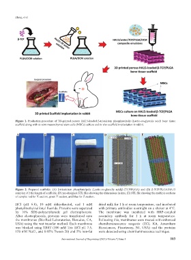
Figure 1. Production procedure of 3D-printed porous HA15-loaded β-tricalcium phosphate/poly (Lactic-co-glycolic acid) bone tissue
scaffold along with in vitro mesenchymal stem cells (MSCs) culture and in vivo scaffold implantation in rabbit.
A B C D E
Figure 2. Prepared scaffolds. (A) β-tricalcium phosphate/poly (Lactic-co-glycolic acid)β-(TCP/PLGA) and (B) β-TCP/PLGA/HA15
samples; (C) the length of scaffolds; (D) pre-designed STL files showing the dimensions in mm; (E) STL file showing the multiple sections
of sample: red for X section, green Y section, and blue for Z section.
HCl (pH 6.8), 10 mM dithiothreitol, and 1 mM dried milk for 1 h at room temperature, and incubated
phenylmethylsulfonyl fluoride. Proteins were separated with primary antibodies overnight on a shaker at 4°C.
by 10% SDS-polyacrylamide gel electrophoresis. The membrane was incubated with HRP-coupled
After electrophoresis, proteins were transferred onto secondary antibody for 1 h at room temperature.
the membranes (Bio-Rad Laboratories, Hercules, CA, Following this, membranes were treated with enhanced
USA) using the wet transfer method. Each membrane chemiluminescence reagents (ECL Kit, Amersham
was blocked using TBST (100 mM Tris–HCl pH 7.5, Biosciences, Piscataway, NJ, USA) and the proteins
150 mM NaCl, and 0.05% Tween 20) and 5% non-fat were detected using chemiluminescence technique.
International Journal of Bioprinting (2021)–Volume 7, Issue 1 103