
Page 79 - IJB-7-1
P. 79
Jing, et al.
A B C D E
F G H I J
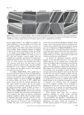
Figure 8. Fiber surface morphological changes of before and after the leaching treatment for 48 h. (A) and (F) poly(ε-caprolactone) (PCL).
(B) and (G) PCL/zein-10. (C) and (H) PCL/zein-20. (D) and (I) PCL/gliadin-10. (E) and (J) PCL/gliadin-20. PCL/gliadin and PCL/zein
scaffolds were leached in culture media and 70% ethanol, respectively. (PCL scaffold was used as control).
of their amphiphilicity . It is difficult to compare the for the PCL/zein-20 and PCL/gliadin-20 scaffolds, which
[17]
cellular behaviors of zein and gliadin on PCL/zein and is due to the increasing portion and size of plant protein
PCL/gliadin scaffolds in the same way, as gliadin can particles in the composite inks. For example, the average
quickly dissolve in the culture medium but zein has poor pore size of nanopores on Figure 8I and 8J is 133.1 ±
water solubility. Hence, leaching treatment was applied 47.4 nm and 209.2 ± 76.2 nm, respectively. These holes
to simulate the surface morphology change of scaffolds and fissures interconnected with each other, generated a
in vitro. Before the leaching treatment, the PCL, PCL/ highly cavernous structure, and increased surface area
zein, and PCL/gliadin scaffolds were dried in vacuo at exponentially; all of which significantly facilitated cell
40°C until they reached a constant weight. Then, the migration, proliferation, and infiltration.
scaffolds were completely immersed in 70% ethanol with In general, the developed composite scaffolds
shaking (80 rpm) for 48 h. The solution was replenished degrade in two interrelated processes. First, zein and
every 12 h. After treatment, the scaffolds were washed gliadin particles can be released from scaffold fiber
with deionized water thrice and then dried in vacuo surface under physiological environment, but in different
until they reached a constant weight. Using this method, ways. Since gliadin is a water-soluble protein, its
nanoparticles on the composite scaffold fiber surface particles from PCL/gliadin scaffolds can be dissolved in
were released into the culture medium, and nanopores PBS solution. For PCL/zein scaffolds, various proteases
and cracks were generated. can hydrolyze zein into peptides or amino acids. Thus,
According to Figure 8A-E, surface morphology of nanopores and cracks on the fiber surface can be observed
fibers was almost the same for all the scaffold materials on both PCL/zein and PCL/gliadin scaffolds. Second,
because relatively small portion of nanoparticles in the such nanoporous surface can accelerate the composite
composite ink materials was not sufficient to engender scaffolds’ degradation, since PCL polymer chains might
noticeable changes. As shown in Figure 8A and 8F, be synchronously released into solution when zein or
no obvious change was found on PCL scaffold fiber gliadin domain in the composite is degraded as a result
surface before and after the leaching treatment since of molecular level blending. Thus, the degradation
PCL cannot dissolve in ethanol. The size and density rate of the composite scaffolds can be controlled by
of gliadin and zein particles could be speculated from adjusting the mixing ratio of plant protein and PCL in
the surface morphology change of fibers, as shown in the biomaterial ink. In general, the composite material
Figure 8G-J, through leaching. Based on the voids on scaffolds degrade faster than the pure PCL scaffolds with
the fiber surface (Figure 8G-8J), it can be speculated that the same scaffold structural parameters.
both zein and gliadin can self-assemble into nanoparticles Plant proteins have been used to develop
in the composite ink, which can influence mechanical composite scaffolds for several reasons, including
properties of printed scaffolds. Moreover, higher density their biocompatibility, biodegradability, safety, low
of nanopores and cracks could be observed on PCL/ cost, processibility, and ductility . Nevertheless, the
[28]
zein-20 and PCL/gliadin-20 scaffolds compared with possible immunogenicity effects restrict their biomedical
those observed on PCL/zein-10 and PCL/gliadin-10 applications. Of course, whether the occurrence of
scaffolds. The scale of such nanopores was also larger immune response depends on the dose of plant protein
International Journal of Bioprinting (2021)–Volume 7, Issue 1 75