
Page 77 - IJB-7-1
P. 77
Jing, et al.
A
B
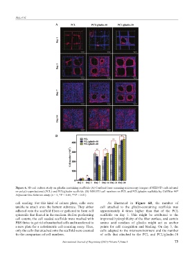
Figure 6. 3D cell culture study on gliadin containing scaffolds (A) Confocal laser scanning microscopy images of NIH/3T3 cell cultured
on poly(ε-caprolactone) (PCL) and PCL/gliadin scaffolds. (B) NIH/3T3 cell numbers on PCL and PCL/gliadin scaffolds by CellTiter 96
®
AQueous One Solution assay (n = 5, *P < 0.05, **P < 0.01).
cell seeding. For this kind of culture plate, cells were As illustrated in Figure 6B, the number of
unable to attach onto the bottom substrate. They either cell attached to the gliadin-containing scaffolds was
adhered onto the scaffold fibers or gathered to form cell approximately 4 times higher than that of the PCL
spheroids that floated in the medium. Before performing scaffolds on day 1. This might be attributed to the
cell counts, the cell seeded scaffolds were washed with improved hydrophilicity of the fiber surface, and certain
PBS thrice to get rid of unattached cells and transferred to amino acid residues of gliadin might act as anchor
a new plate for a colorimetric cell counting assay. Thus, points for cell recognition and binding. On day 3, the
only the cells that attached onto the scaffold were counted cells adapted to the microenvironment and the number
for the comparison of cell numbers. of cells that attached to the PCL and PCL/gliadin-10
International Journal of Bioprinting (2021)–Volume 7, Issue 1 73