
Page 76 - IJB-7-1
P. 76
Using Plant Proteins to Develop Composite Scaffolds
degrades at a proper rate during tissue regeneration (2) Cell viability and proliferation studies in the
process. This is not a serious issue for cell culture studies PCL/gliadin scaffolds
since scaffolds are only used for a short time to produce The cellular interactions of these fabricated
in vitro models. PCL/gliadin scaffolds were further studied in this
A well-known cell line derived from mouse embryo sub-section. The influence of gliadin component was
cells, named NIH/3T3 cell, was cultured on PCL, investigated by directly culturing NIH/3T3 cells on the
PCL/gliadin, and PCL/zein scaffolds for biocompatibility PCL/gliadin scaffolds. The scaffolds were cut into unified
evaluation. Due to the concern of side effects from gliadin round specimens and inserted in the ultralow attachment
in cell growth, the cytotoxicity of PCL/gliadin scaffolds culture plate. A small volume of cell suspension was
was evaluated before cell culture studies. directly pipetted onto the specimen in each well for cell
seeding. After incubation for few hours, the culture medium
(1) Cytotoxicity assay of gliadin released from was added, and some cells adhered onto the scaffold fibers.
PCL/gliadin scaffolds Cell seeded scaffolds were visualized by confocal
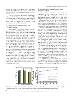
The cytotoxicity of the gliadin released from PCL/ laser scanning microscopy (CLSM, LSM-880, ZEISS,
gliadin scaffolds on NIH/3T3 fibroblast cell was evaluated Germany). After fixation in paraformaldehyde, the cell
through colorimetric MTT assay. As shown in Figure 5A, nuclei and membrane were stained with Hoechst 33342
the cell viability of NIH/3T3 cells was higher than 95% and DiI dye, which emit blue and red fluorescence,
when incubating together with PCL or PCL/gliadin-10 respectively, on laser excitation. Figure 6A shows the
scaffolds. The cell viability for PCL/gliadin-10 scaffolds CLSM images of cell cultured on PCL and PCL/gliadin
was slightly higher than that of the control group (cell scaffolds on days 1, 3, 7, and 14. On day 1, the spheroid
culture in medium), while this value was only about 85% cells could partially attach onto the scaffolds. After 3
days, most of the attached cells started to spread, orient,
for PCL/gliadin-20 scaffolds. This suppressive effect elongate along the fiber directions, and extend the
was caused increased release of gliadin from the PCL/ filopodia at leading edge to grab on the adjacent fibers.
gliadin-20 scaffold since its corresponding weight loss in This resembles fibroblast migration in vivo with front-end/
72 h was obviously bigger than that of PCL/gliadin-10. back-end polarity . The cell adhesion and proliferation
[27]
The weight loss of PCL/gliadin scaffolds was were not homogeneous on the first few days because of
measured by immersing them in phosphate-buffered saline uneven cell distribution during initial cell seeding. From
(PBS, 10 mM, pH 7.4) with the addition of 1% antibiotics day 3 to day 7, the fibroblasts actively proliferated from
at an incubator (37°C). As in Figure 5B, the weight the scaffold’s side walls to the center of pores, formed a
loss of PCL scaffold versus time was negligible, while unique circular structure, and eventually merged to form
the weight loss for PCL/gliadin-10 and PCL/gliadin-20 a cellular film. Finally, a film-like cell sheet was observed
scaffolds was up to 14.5% and 21.2%, respectively, in in each cultured scaffold on day 14.
120 h. Nearly 90% of the total weight loss happened in A colorimetric cell proliferation assay (CellTiter
the first 20 h. Meanwhile, the release of gliadin did not 96 AQueous One Solution) was applied to count cell
®
change the pH value of the solution as gliadin is neutral numbers. In cell culture experiments, the ultra-low
substance in the solution. attachment culture plate (Corning 3473) was used for
A B
Figure 5. In vitro cytotoxicity test (A) Cell viability and relative dry mass loss after seeding cells on poly(ε-caprolactone) (PCL) and
PCL/gliadin scaffolds for 72 h; (B) weight loss profile of PCL and PCL/gliadin scaffolds in phosphate-buffered saline.
72 International Journal of Bioprinting (2021)–Volume 7, Issue 1