
Page 89 - IJB-7-1
P. 89
Xie, et al.
A B
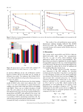
Figure 9. Number of colony-forming units/mL of Staphylococcus aureus after incubation with different samples at (A) uncontrolled pH
values and (B) neutral pH of 7.4.
The results of the cell proliferation assays and the
fluorescence staining imaging indicated that SLMed
ZK30-0.2Cu-xMn has suitable cytocompatibility as
expected, because all elements in the SLMed alloys are
cytocompatible.
4. Discussion
This current study successfully used SLM to fabricate
ZK30-0.2Cu-xMn with various Mn concentrations
(0.4, 0.8, 1.2, and 1.6 wt%), which exhibited strong
antibacterial ability and good cytocompatibility. Mg-
based alloys are attractive candidates for metallic implant
biodegradable materials if the rate of the biodegradation
can be decreased. The decrease of the biodegradation rate
was examined by adding Mn by SLM for antibacterial
Figure 10. Optical density values for MG63 cells incubated in Ti Cu-containing Mg alloys in this study.
and SLMed ZK30-0.2Cu-xMn extracts for 1, 4, and 7 days. After the SLMed ZK30-0.2Cu-xMn specimens
were immersed in the SBF solution, the corrosion
no apparent difference in the cell proliferation number reactions given by Equation 4, Equation 5, and Equation
between SLMed ZK30-0.2Cu-xMn and Ti extracts at 6 in the following occurred and the surface hydroxide
different time points. This indicates that SLMed ZK30- layer formed. The hydroxide layer was loose (Figure 7A)
0.2Cu-xMn is suitable for cell growth without cytotoxicity and provided little corrosion protection. As corrosion
as well as Ti extract. continued, hydroxyapatite formed due to the reaction
2-
Figure 11 shows the fluorescence live/dead staining between hydroxide in the corrosion product, HPO
4
2+
results of MG63 cells, which were cultured after 1 and (or PO ) and Ca in the SBF, which resulted in the
3-
4
3 days. Live cells were indicated by green fluorescence precipitation of Ca/P hydroxide on the surface of the
[32]
staining with calcein-AM, while dead cells by red staining hydroxide layer according to Equation vii . This was
with EthD-1. There was no evident indication of dead substantiated by the EDS spectra shown in Figure 7F,
cells. The cells cultured on all the SLMed alloys had a which indicated that the corrosion products are mainly
well spread morphology, exhibiting no apparent difference composed of O, Mg, Ca, and P.
among them and MG63 cells cultured in the extract had Mg − 2e → Mg 2+ (iv)
2+
−
a morphology with spindle and round shapes which was
in accordance with the morphology of normal cells. The 2H O + O + 4e → 4OH − (v)
−
cells showed good growth patterns and the number of cells 2 2
increased obviously as incubation time increased. Mg + 2OH →Mg(OH) 2 (vi)
2+
−
International Journal of Bioprinting (2021)–Volume 7, Issue 1 85