
Page 114 - IJB-7-2
P. 114
Technique of Thyroid Cartilage Scaffold Support Formation
A B on the design of the study, internal structure and geometry
of a scaffold, the polymerization and/or cross-linking occur
before or during bioprinting, or even after the formation
of the whole scaffold. The guided polymerization and/
or cross-linking of the biomaterial are influenced by
different physical and chemical factors: UV, temperature,
ion concentration, pH, etc. Collagen is a fibrillar protein
comprising of connective tissue (tendon, bone, cartilage,
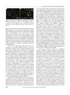
Figure 7. Estimation of viability rat chondrocytes in collagen dermis, etc.). Collagen type I is characterized by high
scaffold through staining (A) On the 3 and (B) 7 days of affinity to the cells and lack of species specificity [1,30] .
th
rd
incubation. The green indicates live cells, while the red indicates The material provides cell adhesion, migration,
dead cells. Composite images were made of 10 layers. Scale bar – proliferation, and differentiation. As a component of
100 μm. extracellular matrix, collagen ensures the rigidity and
integrity of a tissue . However, collagen itself has weak
[2]
This method provides well-formed porous structures, mechanical properties and a high rate of biodegradation.
large surface area and space for cell attachment, growth, The native way of collagen polymerization is a shift in
and proliferation. Moreover, larynx and trachea scaffolds temperature (increase) and pH (increase) . Another
[31]
made of polypropylene mesh covered with collagen option suggests inverse pH change (decrease), but it
sponge have already been used clinically . The will result in low cell viability [32,33] . The applicability of
[19]
technology involves the use of a tube-shaped frame (made additional chemical cross-linking with genipin , tannic
[34]
of polypropylene mesh), as well as polypropylene support acid , glutaraldehyde , and dialdehyde starch
[35]
[37]
[36]
ring embedded in it to provide rigidity to the structure. has been shown. Drzewiecki et al. described the use of
The inner and outer surfaces of the tubes were coated with photocuring collagen methacrylamide as a bio-ink (cell-
collagen. Our results on collagen biocompatibility testing laden biomaterial applicable for bioprinting) . Another
[38]
confirm suitability of collagen as a substrate/scaffold for option is riboflavin that provides the formation of covalent
cell proliferation. crosslinks in collagen . In the present study, no forced
[39]
Since both technologies described above are cross-linking and high concentration of the material were
related to the acellular scaffold type, they could not used to increase stiffness of the scaffold.
be directly compared with the method described in the In our study, gelatin served as the supporting
study. The technique which is closest to our approach material. Gelatin is a collagen hydrolysate. and has the
was described by Hinton et al. . They used freeform same amino acid composition . Unlike collagen, gelatin
[20]
[40]
reversible embedding of suspended hydrogels (FRESH). is soluble in water. In tissue engineering, gelatin is used as
Based on the technique, printing hydrogel was embedded sacrificial material that could be easily removed at 37°C
in a secondary hydrogel, which serves as temporary (standard temperature for cell culture and tissue scaffolds
thermoreversible and biocompatible support. 3D-imprints incubation). In general, gelatin is not characterized
of alginate, fibrin, matrigel, and collagen were obtained. by antigenic and immunogenic properties . It is
[41]
The method was later improved to ensure higher relatively cheap, making it a very attractive material
printing accuracy . Other similar techniques have been for 3D-bioprinting. For many years, its reversible “Sol-
[21]
described by Bhattacharjee et al. and Wu et al. . Gel” transition has been used for temporal (sacrificial)
[22]
[23]
An alternative approach for biofabrication of a scaffold scaffold elements. Recently, the use of a commercial
with high complexity in its geometry is related to the product called Pluronic F-127 has become extensive for
use of reverse strategy: Placement of sacrificial material this purpose [31,42,43] . The material was also adapted to form
in main material. Basically, such constructions serve as macropores in a scaffold . Fitzsimmons et al. showed
[31]
[42]
an additional means of forming hollow channels [24-28] . that Pluronic F-127 is superior to gelatin as a sacrificial
However, when the geometry is complicated, the use of material for creating vascularized tissues. Both gelatin
an additional custom-shaped mold may be required in and Pluronic F-127 have high printability and ease of
order to form the scaffold, and bioprinting is used only to removal. Filament printed with Pluronic F-127 has higher
create the internal structure of the scaffold . spatial resolution, greater uniformity, and modulus of
[29]
Most of the biomaterials used for bioprinting are elasticity than the one of gelatin . At the same time, the
[42]
usually soft (resulting in fragility of the printed scaffolds); use of Pluronic F-127 strictly requires incubation at low
therefore, there is a high probability of deformation of the temperature (e.g., 4°C) after the printing to complete its
material under its own weight. For the formation of an removal from a scaffold , while gelatin can easily melt at
[44]
elastic filament during the material extrusion, a stage of standard temperature (37°C). Gelatin could be considered
polymerization and/or cross-linking is urgent. Depending as a more affordable material and thus, the first choice for
110 International Journal of Bioprinting (2021)–Volume 7, Issue 2