
Page 13 - IJB-7-2
P. 13
Agung, et al.
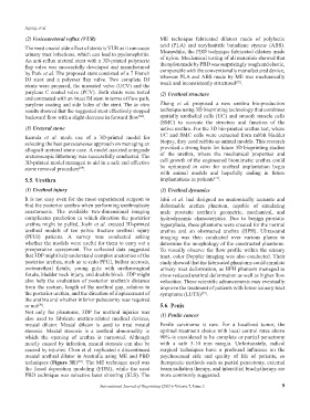
(2) Vesicoureteral reflux (VUR) ME technique fabricated dilators made of polylactic
acid (PLA) and acrylonitrile butadiene styrene (ABS).
The most crucial side effect of stents is VUR as it can cause Meanwhile, the PBD technique fabricated dilators made
urinary tract infections, which can lead to pyelonephritis.
An anti-reflux ureteral stent with a 3D-printed polymeric of nylon. Mechanical testing of all materials showed that
flap valve was successfully developed and manufactured the nylon made by PBD was surprisingly tough and elastic,
by Park et al. The proposed stent consisted of a 7 French comparable with the conventionally manufactured device,
DJ stent and a polymer flap valve. Two complete DJ whereas PLA and ABS made by ME was mechanically
[72]
stents were prepared, the uncoated valve (UCV) and the weak and inconsistently structured .
parylene C coated valve (PCV). Both stents were tested (2) Urethral structure
and contrasted with an intact DJ stent in terms of flow path,
parylene coating and side holes of the stent. The in vitro Zhang et al. proposed a new urethra bio-production
results showed that the suggested stent effectively stopped technique using 3D bioprinting technology that combines
backward flow with a slight decrease in forward flow . spatially urothelial cells (UC) and smooth muscle cells
[68]
(SMC) to recreate the structure and function of the
(3) Ureteral stone native urethra. For the 3D bio-printed urethra test, where
Kuroda et al. made use of a 3D-printed model for UC and SMC cells were extracted from rabbit bladder
selecting the best percutaneous approach on managing an biopsy, they used rabbits as animal models. This research
allograft ureteral stone case. A model-assisted antegrade provided a strong basis for future 3D bioprinting studies
ureteroscopic lithotomy was successfully conducted. The of the urethra, where the mechanical properties and
3D-printed model managed to aid in a safe and effective cell growth of the engineered biomimetic urethra could
stone removal procedure . be optimized in vitro for urethral implantation begin
[69]
with animal models and hopefully ending in future
5.5. Urethra implantations in patients .
[71]
(1) Urethral injury (3) Urethral dynamics
It is not easy even for the most experienced surgeon to Ishii et al. had designed an anatomically accurate and
find the posterior urethra when performing urethroplasty deformable urethra phantom, capable of simulating
anastomosis. The available two-dimensional imaging male prostatic urethra’s geometric, mechanical, and
complicates prediction in which direction the posterior hydrodynamic characteristics. Due to benign prostatic
urethra might be pulled. Joshi et al. created 3D-printed hyperplasia, these phantoms were created for the normal
urethral models of ten pelvic fracture urethral injury urethra and an obstructed urethra (BPH). Ultrasound
(PFUI) patients. A survey was conducted asking imaging was then conducted over various planes to
whether the models were useful for them to carry out a determine the morphology of the constructed phantoms.
preoperative assessment. The collected data suggested To visually observe the flow profile within the urinary
that 3DP might help understand complex anatomies of the tract, color Doppler imaging was also conducted. Their
posterior urethra, such as re-redo PFUI, bulbar necrosis, study showed that the fabricated phantoms could simulate
rectourethral fistula, young girls with urethrovaginal urinary tract deformation, as BPH phantom managed to
fistula, bladder neck injury, and double block. 3DP might show reduced urethral deformation as well as higher flow
also help the evaluation of posterior urethra’s distance velocities. These scientific advancements may eventually
from the rectum, length of the urethral gap, relation to improve the treatment of patients with lower urinary tract
the posterior urethra, and the direction of displacement of symptoms (LUTS) .
[27]
the urethra and whether inferior pubectomy was required
or not . 5.6. Penis
[70]
Not only for phantoms, 3DP for urethral injuries was (1) Penile cancer
also used to fabricate urethra-related medical devices,
meatal dilator. Meatal dilator is used to treat meatal Penile carcinoma is rare. For a localized tumor, the
stenosis. Meatal stenosis is a urethral abnormality in optimal treatment choice with local control rates above
which the opening of urethra is narrowed. Although 90% is considered to be complete or partial penectomy
mostly caused by infection, meatal stenosis can also be with a safe 5-10 mm margin. Unfortunately, radical
caused by injuries. Chen et al. replicated a discontinued surgical techniques have a profound influence on the
meatal urethral dilator in Australia using ME and PBD psychosexual side and quality of life of patients, so
techniques (Figure 3E) . The ME technique used was therapeutic methods such as partial penectomy, external
[72]
the fused deposition modeling (FDM), while the used beam radiation therapy, and interstitial brachytherapy are
PBD technique was selective laser sintering (SLS). The more commonly suggested.
International Journal of Bioprinting (2021)–Volume 7, Issue 2 9