
Page 6 - IJB-7-2
P. 6
The Role of 3DP Phantoms and Devices for Organ-specified Appliances in Urology
3D-printed models were shown to be superior to 3D images contoured in an xy-plane before finally, the layer stack
in pre-operative planning. This was due to the ability of 3D forms the third (z) dimension. In 2015, the ISO/ASTM
models to provide more details and tactile representations of 52900 Standard was set and established seven categories
organ anatomical aspects for the operators [13-15] . in the 3DP processes. These categories include material
The anatomical models may also be used for mock extrusion (ME), vat polymerization (VP), powder bed
surgeries and pre-surgical adjustment of instrumentation, fusion (PBD), material jetting (MJ), binder jetting (BJ),
thus reducing the operation time, ensuring better approach, direct energy deposition (DEP), and sheet lamination
and instrument compatibility. Several surgeries have (SL). Each process has its supporting technology as well
received advantages from this approach, including vascular as the materials used [21,22] .
surgery for endovascular aneurysm repair, cardiac surgery Each category of 3DP processes in the ISO/ASTM
for pre-surgical tumor resection planning and congenital 52900 Standard has also been used for medical purposes .
[23]
defect repair, neurosurgery for navigation training, and ME and VP have been used for casing fabrication of medical
in orthopedic surgery for tumor resection planning and instruments [7,24] , scaffolds , prostheses [25,26] , and phantoms
[6]
[27]
trauma injury treatment . In addition to its utilization as of organs . Meanwhile, PBD, MJ, BJ, DEP, and SL are
[15]
an anatomical model and surgical guide, 3DP technology reported to be used for bone reconstructions and porous
is also commonly used in implant manufacturing. Patient- implants made of metals (i.e., Fe, Mg, and Ti) [28,29] . Metal-
specific implant (PSI) is a perfect-fit implant used to restore based scaffolds can also be fabricated using MJ and BJ ,
[29]
the anatomy, relationship, and function of a patient’s organ. which are mainly applied for bone fracture repairment.
These implants have been reported in orthopedics, thoracic Printing a 3D printed model requires several steps
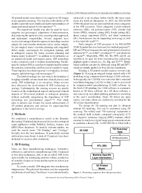
surgery, ophthalmology, and neurosurgery . (Figure 1). Printing an original design started with a 3D
[15]
The field of urology has also been at the frontline of modeling using computer-aided design (CAD) software.
bringing scientific advancement into clinical practice and Subsequently, the CAD file was converted into a standard
surely 3DP technology is no exception. Many reviews tessellation language (.stl) file so that it can be processed
have discussed the involvement of 3DP technology in in computer-aided manufacturing (CAM) software. In
urology. Unfortunately, the existing reviews are mostly the field of 3D printing, the CAM software is commonly
focused on the technological aspects and general clinical known as 3D slicer software. In a 3D slicer software, a
impacts of 3D-printed products in urological practices user can set up and adjust printing parameters according
without explicitly categorizing the importance of 3DP to the user’s specification. Some 3D slicers are open
technology per genitourinary organ [16-20] . This study source, but some others are exclusively provided by the
aims to present and discuss the recent advancement of 3D printer manufacturers.
3D printed phantoms and devices for organ-specified The design for 3D printing can also be obtained
appliances in the field of urology. without 3D modeling. This can be performed using 3D
scanning. The widely used “3D scanning” equipment in
2. Methods the medical field includes computed tomography (CT)
and magnetic resonance imaging (MRI) scanners, which
We conducted a comprehensive search in the literature
discussing 3D printed phantoms and devices for urological commonly results in a digital imaging and communications
organ appliances within PubMed, Embase, Scopus, and in medicine (.dicom) file. A DICOM file is filled with a
EBSCOhost databases. To identify relevant studies, we
used the search terms “3D Printing” and “Urology.”
Initially, from the four databases, 56 potentially relevant
publications were listed. A total of 35 journals have been
included for analysis after exclusions.
3. 3D Printing
There are three manufacturing techniques: Formative,
subtractive, and additive manufacturing (AM). Compared
to two other techniques, AM, or the so-called 3DP, is the
most suitable for prototyping and low volume production
of complex designs as it may produce parts in almost any
geometry .
[21]
To create a model, 3DP integrates two simultaneous
subprocesses: The physical formation and the sequential
attachment of each layer. Each layer is two-dimensionally Figure 1. 3D printing sequences from design to product.
2 International Journal of Bioprinting (2021)–Volume 7, Issue 2