
Page 7 - IJB-7-2
P. 7
Agung, et al.
two-dimensional (2D) image of a scanned body part. concluded that pre-operative 3D-printed models improved
The compilation of DICOM files for a body part can be patients’ understanding of their condition and the goals
reconstructed into a 3D model , which is subsequently of the surgery . Similar findings to Wake et al., Ilie
[30]
[35]
converted into a standard tessellation (.stl) file for 3D et al. demonstrated the questionnaire data that show
printing . However, before conversion, the 3D model is the satisfaction of the patients regarding the use of
[31]
refined or segmented to isolate a specific to-print tissue. The 3D-printed model during the clinical case discussion and
refinement is required since the 3D reconstructed model were satisfied with this new way of communication .
[36]
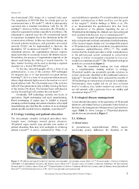
is sometimes incomplete due to the limitations in the 3D On the other hand, 3D-printed organ models may also
reconstruction software. In this case, transfer learning, one help urologists and residents understand detailed anatomy
of neural network techniques, and generative adversarial of the diseased organs. Atalay et al. investigated the impact
network (GAN) can be implemented to fine-tune the of 3D-printed renal models on residents’ perception before
incomplete 3D reconstructed model [32,33] . Similar to the percutaneous nephrolithotomy (PNL) [37] . The results
refinement process, the segmentation process requires showed that the models provided a better comprehension
neural networks to accurately classify the desired tissues. of the pelvicalyceal system compared to conventional
Nonetheless, the accuracy of segmentation depends on the imaging. A similar investigation conducted by Lee et al.
dataset used during the training of neural networks. To resulted in consistent results . The 3D printed urological
[38]
date, transfer learning can be used to develop a segment models are presented in Figure 2.
classifier for a limited DICOM data . Since the simulation training has been utilized
[32]
To this day, stereolithography (SLA), a form of vat as a complementary training method in urology,
polymerization, is one of the common 3DP techniques 3D-printed models can potentially provide solutions to
for surgeries due to its high precision and great surface several drawbacks identified in the traditional cadaveric
finishing . SLA is a form of vat polymerization process training . Several studies have indicated the benefits of
[34]
[39]
where a high intensity light source is focused on to a vat of 3DP technology in various types of urological simulations.
liquid polymer bath. The illuminated area of the polymer Unfortunately, these studies did not yet have sufficient
bath will thus photochemically solidify, forming the layer level of evidence, thus further randomized control trials
of the desired 3D object. The finished layer will descend are still needed, with a particular focus on validity and
and the focused light will renders the next layer . educational impact [16,40-45] .
[34]
Eventually, 3DP technology provides two levels of
application: Rapid prototyping and rapid manufacturing. 5. Urological disease management
Rapid prototyping of organs may be helpful in surgical A more detailed description of the application of 3D-printed
planning, resident training, and patient education, while rapid
manufacturing may facilitate the creation of an on-demand phantoms and related devices is presented below based on
patient-specific medical devices, implants, or prostheses . their use in the management of diseases in each genitourinary
[22]
organ (Table 1). Some footages of 3D-printed phantoms
4. Urology training and patient education and devices are presented in Figure 3.
The increasingly complex urological procedures have 5.1. Kidneys
brought more challenges toward patient education. (1) Renal stones
3D-printed organ models may provide new modes of
patient education, thus may help urologists in obtaining In percutaneous nephrolithotomy (PNL), needle
patient consent . A survey conducted by Wake et al. positioning greatly influences the duration of the
[34]
A B C D
Figure 2. 3D printed urological models for training and education: (A) prostate cancer (from ref. licensed under a Creative Commons
[35]
Attribution 4.0 International License), (B) kidney cancer (from ref. licensed under a Creative Commons Attribution 4.0 International
[35]
License) and (C) kidney cancer (from ref. licensed under a Creative Commons Attribution License), and (d) kidney stone [reproduced
[38]
from ref. with the kind permission of Dr. Lütfi Canat (private communication)].
[47]
International Journal of Bioprinting (2021)–Volume 7, Issue 2 3