
Page 94 - IJB-7-3
P. 94
Coaxial Electrohydrodynamic Bioprinting of Pre-Vascularized Tissues
A B C
D E
F G H
I J K
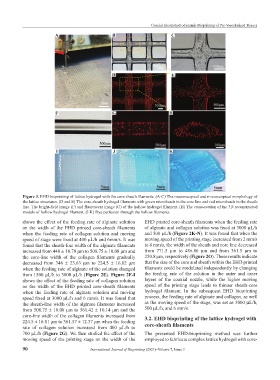
Figure 3. EHD bioprinting of lattice hydrogel with the core-sheath filaments. (A-C) The macroscopical and microscopical morphology of
the lattice structures. (D and E) The core-sheath hydrogel filaments with green microbeads in the core line and red microbeads in the sheath
line. The bright-field image (F) and fluorescent image (G) of the hollow hydrogel filament. (H) The cross-section of the 3D reconstructed
models of hollow hydrogel filament. (I-K) Dye perfusion through the hollow filaments.
shows the effect of the feeding rate of alginate solution EHD printed core-sheath filaments when the feeding rate
on the width of the EHD printed core-sheath filaments of alginate and collagen solution was fixed at 3000 μL/h
when the feeding rate of collagen solution and moving and 500 μL/h (Figure 2K-N). It was found that when the
speed of stage were fixed at 400 μL/h and 6mm/s. It was moving speed of the printing stage increased from 2 mm/s
found that the sheath-line width of the alginate filaments to 8 mm/s, the width of the sheath and core line decreased
increased from 440 ± 18.79 μm to 508.75 ± 10.08 μm and from 771.5 μm to 486.86 μm and from 361.5 μm to
the core-line width of the collagen filaments gradually 250.8 μm, respectively (Figure 2O). These results indicate
decreased from 346 ± 23.63 μm to 224.5 ± 16.82 μm that the size of the core and sheath within the EHD printed
when the feeding rate of alginate of the solution changed filaments could be modulated independently by changing
from 1500 μL/h to 3000 μL/h (Figure 2E). Figure 2F-I the feeding rate of the solution in the outer and inner
shows the effect of the feeding rate of collagen solution layers of the coaxial nozzle, while the higher moving
on the width of the EHD printed core-sheath filaments speed of the printing stage leads to thinner sheath-core
when the feeding rate of alginate solution and moving hydrogel filament. In the subsequent EHD bioprinting
speed fixed at 3000 μL/h and 6 mm/s. It was found that process, the feeding rate of alginate and collagen, as well
the sheath-line width of the alginate filaments increased as the moving speed of the stage, was set as 3000 μL/h,
from 508.75 ± 10.08 μm to 561.42 ± 10.14 μm and the 500 μL/h, and 6 mm/s.
core-line width of the collagen filaments increased from 3.2. EHD bioprinting of the lattice hydrogel with
224.5 ± 16.81 μm to 361.57 ± 12.37 μm when the feeding core-sheath filaments
rate of collagen solution increased from 400 μL/h to
700 μL/h (Figure 2G). We then studied the effect of the The presented EHD-bioprinting method was further
moving speed of the printing stage on the width of the employed to fabricate complex lattice hydrogel with core-
90 International Journal of Bioprinting (2021)–Volume 7, Issue 3