
Page 96 - IJB-7-3
P. 96
Coaxial Electrohydrodynamic Bioprinting of Pre-Vascularized Tissues
A B C D
E F G H
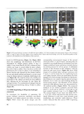
Figure 5. 3D porous hydrogel constructs with different layer numbers. (A-C) Macroscopical and microscopical images of the constructs
with 3, 5, and 10 layers. (D) SEM images of the constructs with 5 layers after freeze-drying. (E-G) The 3D confocal profiles of the
constructs. (H) Quantification of the thickness of the constructs.
bioink for EHD-bioprinting (Figure 4A). Figure 4B-D corresponding microscopical images of the printed
shows the morphology and distribution of HUVECs constructs with 3, 5, and 10 layers, indicating that the
encapsulated in collagen gelation during 14 days in assembled core-sheath filaments maintained good line
culture. It was found that the HUVECs maintained high feature, predefined layout, and lattice structures during
viability and began to spread within the inner collagen the printing process for all the groups. A substantial
layer after 2 days in culture. There was then a notable cell increase in the thickness of the constructs could be
proliferation after 14 days in culture. It was interesting to observed with more layers. Figure 5D shows SEM
observe that the cells migrated into the interface between images of the printed lattice hydrogel constructs with
the core and sheath hydrogel and formed a circular vessel 5 layers. It can be found that the neighboring layers
along the filament instead of randomly distributing in the were tightly merged, which still maintained structural
core (Figure 4E). This might be owing to the swelling and integrity after freeze-drying. The diameter of the freeze-
degradation of the collagen gel during the long culturing dried filaments was smaller than the printed hydrogel
time, allowing for the movement of cells within the filaments due to water loss and shrinking during the
hydrogel filament. The endothelialized hydrogel filaments freeze-drying process. The thickness of the printed
maintained good morphology during 14 days in culture constructs with different layers was characterized by
(Figure 4F). 3D confocal profiles (Figure 5E-G). The measured
thickness of the printed constructs increased from
3.4. EHD-bioprinting of 3D porous hydrogel 894.35 ± 41.50 μm to 2893.85 ± 92.60 μm as the
constructs layer number increased from 3 to 10 (Figure 5H).
The average height of each layer was about 290.75
To investigate the feasibility of generating 3D ± 12.22 μm, which indicated that the printed core-
porous hydrogel constructs, multilayer core-sheath sheath filaments in each layer could maintain good
filaments were further printed into lattice structures. morphology and relatively stable thickness. These
Figure 5A-C shows the photo images and the results indicated that this coaxial EHD bioprinting
92 International Journal of Bioprinting (2021)–Volume 7, Issue 3