
Page 95 - IJB-7-3
P. 95
Mao, et al.
A
B
C
D
E F
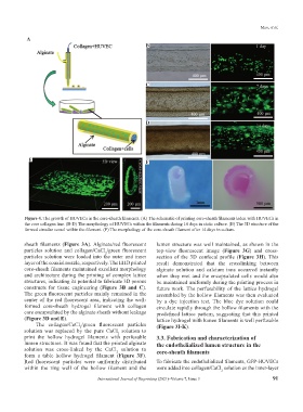
Figure 4. The growth of HUVECs in the core-sheath filaments. (A) The schematic of printing core-sheath filaments laden with HUVECs in
the core collagen line. (B-D) The morphology of HUVECs within the filaments during 14 days in static culture. (E) The 3D structure of the
formed circular vessel within the filament. (F) The morphology of the core-sheath filament after 14 days in culture.
sheath filaments (Figure 3A). Alginate/red fluorescent lumen structure was well maintained, as shown in the
particles solution and collagen/CaCl /green fluorescent top-view fluorescent image (Figure 3G) and cross-
2
particles solution were loaded into the outer and inner section of the 3D confocal profile (Figure 3H). This
layer of the coaxial nozzle, respectively. The EHD printed result demonstrated that the crosslinking between
core-sheath filaments maintained excellent morphology alginate solution and calcium ions occurred instantly
and architecture during the printing of complex lattice when they met and the encapsulated cells would also
structures, indicating its potential to fabricate 3D porous be maintained uniformly during the printing process in
constructs for tissue engineering (Figure 3B and C). future work. The perfusability of the lattice hydrogel
The green fluorescent particles mainly remained in the assembled by the hollow filaments was then evaluated
center of the red fluorescent area, indicating the well- by a dye injection test. The blue dye solution could
formed core-sheath hydrogel filament with collagen circulate rapidly through the hollow filaments with the
core encapsulated by the alginate sheath without leakage predefined lattice pattern, suggesting that this printed
(Figure 3D and E). lattice hydrogel with lumen filaments is well perfusable
The collagen/CaCl /green fluorescent particles (Figure 3I-K).
2
solution was replaced by the pure CaCl solution to
2
print the hollow hydrogel filaments with perfusable 3.3. Fabrication and characterization of
lumen structures. It was found that the printed alginate the endothelialized lumen structure in the
solution was cross-linked by the CaCl solution to core-sheath filaments
2
form a table hollow hydrogel filament (Figure 3F).
Red fluorescent particles were uniformly distributed To fabricate the endothelialized filaments, GFP-HUVECs
within the ring wall of the hollow filament and the were added into collagen/CaCl solution as the inner-layer
2
International Journal of Bioprinting (2021)–Volume 7, Issue 3 91