
Page 193 - IJB-8-1
P. 193
Yao, et al.
series model and each model can be directly trained
under specific cell lines. Another possible way is
to train multiple AD-GANs on different cell lines
independently. When testing on an unseen cell line,
the output of multiple AD-GANs shall be merged or
fused. Moreover, a few aspects of our method should
be further improved, such as network architecture, loss
function design, and data augmentation strategies in
synthetic mask generation. Last but not least, the very
practical dilemma to most researchers is that they may
not be familiar with ML or image processing methods.
It is essential to develop a user-friendly interface so
that researchers without expertise in image analysis
and ML can incorporate 3D nuclei segmentation flow
to perform nuclei classification and cell culture model
analysis.
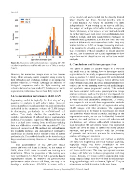
Figure 14. Nuclei size and number analysis of culturing NIH-3T3
on poly-E-caprolactone scaffold with pore size of 100 µm on days 6. Conclusions and future perspectives
3 and 6. The desire to assess 3D culture models in a low-cost
and rapid way is the driving force to investigate nuclei
However, the normalized images more or less become segmentation. In this study, we presented an unsupervised
blurry. More seriously, nuclei elongated along Z-axis by learning method AD-GAN to segment 3D nuclei labeled
light diffraction and scattering, leading to an unexpected with fluorescent in CLSM images, which utilizes both
position offset in 3D voxels. Although the influence of same-domain translation and cross-domain translation to
biological components under the light scattering and achieve one-to-one mapping between real nuclei images
refractive indexes has been studied , their impact on nuclei and synthetic masks (segmented nuclei). This method
[35]
segmentation performance has not been fully explored. has been compared with some general-purpose image
analysis software, such as Cellprofiler and Squassh for
5.2. Generalization performance of AD-GAN 3D nuclei segmentation, and achieves better performance
Segmenting nuclei is typically the first step of any in both visual and quantitative comparison. Of course,
quantitative analysis in cell culture tasks. However, our purpose is not to rank these segmentation methods
extracting subjective and quantitative nuclei information but to evaluate their suitability in cell segmentation using
embedded in the enormous volume of CLSM images CLSM images with fiber structures. The segmented
is challenging, particularly in scaffold-based cell nuclei could help us to bridge the knowledge gap about
culture. This study clarifies the capabilities and cell activities within fibrous scaffolds. Building on the
realistic expectations of different nuclei segmentation segmentation results, we can use the identified the nuclei
methods. For example, supervised ML models typically number, size and position to assess cell adhesion and
face a critical issue coming from insufficient training proliferation, and address cell-scaffold interaction in
data and difficulties related to data annotation [36] . Our high-throughput 3D cell culture model. The segmented
proposed unsupervised ML method has outperformed nuclei can serve as seeds to outline the entire cellular
the available methods and demonstrated comparable structure, and possibly associate with the locations
capabilities to identify nuclei similar to that of human of genomic and proteomic products for morphometry
professionals. The method should also be tested using a analysis of biological structures.
larger database covering diverse cell lines and scaffold We would like to extend our method to analyze
types. cell behaviors in spheroid, tumoroid, hydrogel scaffolds,
The generalization of the AD-GAN model organoids which can better recapitulate in vivo
across different cell lines is limited by the nature of morphology, cell connectivity, polarity, gene expression
GAN, since GAN tends to overfit on training data and tissue architecture. This may open up new avenues
distribution. When the properties of the testing data to study cell behaviors in disease progression and drug
vary significantly, AD-GAN may fail to generate decent release. Besides, we would explore segmentation tasks
segmentation results. To improve the generalization using complex image data derived from the microscopic
performance under diverse cell lines, one way is to imaging technology such as electron, light and X-ray to
develop a multi-modal AD-GAN structure with a obtain more nuclei information in the near future.
International Journal of Bioprinting (2022)–Volume 8, Issue 1 179