
Page 42 - IJB-8-1
P. 42
Controlling Droplet Impact Velocity and Droplet Volume Improves Cell Viability in Droplet-Based Bioprinting
the overall density of the bio-inks generally increases with include the complexity of print-head design such as
increasing cell concentration. The density of the cell-laden inner chamber height and the various types of coating
bio-inks increases from 1007.3 ± 2.6 kg/m at 1 million within the firing chamber. The average cell diameter was
3
cells/mL to 1012. ± 2.2 kg/m at 5 million cells/mL. ~18.2 ± 1.6 µm, and the ratio of nozzle diameter to cell
3
The physical properties (viscosity, surface tension and diameter was ~4.396. It was observed that the cell-laden
density) of the different cell-laden bio-inks were used to bio-ink containing 5 million cells/ml could not be ejected
calculate the dimensionless Z value which helps to predict from the nozzle orifice possibly due to clogging issue.
the printability of a bio-ink (Table 1). The dimensionless Z Hence, the subsequent experiments were conducted using
value is an inverse of the Ohnesorge number (Oh), which can printable cell-laden bio-inks (1 – 4 million cells/mL).
be defined as the ratio between the Reynolds number and the Next, the printed cell output per droplet volume was
square root of the Weber number, and is independent of the evaluated for all the cell-laden bio-inks (1 – 4 million
bio-ink velocity . The viscous dissipation prevents droplet cells/mL) at varying droplet volumes (20 nL, 40 nL and
[15]
formation at low Z values (Z < 2), while undesirable satellite 60 nL). In general, the measured cell output per droplet
droplets form at high Z values (Z > 14) . An increasing volume is less than theoretical number of cells based
[45]
cell concentration generally leads to a lower Z value and the on the cell concentration for all the cell-laden bio-inks
measured Z values of the cell-laden bio-inks in this study (1 – 4 million cells/mL) at all droplet volumes (20 nL,
were within the range of 58.11 (5 million cells/mL) ≤ Z ≤ 40 nL and 60 nL). It is likely that the cells adhere to the
72.92 (1 million cells/mL). The high Z values implied that inner surface of the microchannel wall and accumulate
all the cell-laden bio-inks (1 – 5 million cells/mL) were over time, leading to lower-than-expected cell output.
printable with formation of satellite droplets. Nevertheless, The printed cell output of all the cell-laden bio-inks at
it is important to consider potential clogging of the cell- varying droplet volume was summarized in Figure 2A
laden bio-inks (average cell diameter of ~18.2 ± 1.6 µm) and Table 2. Furthermore, the cell-laden bio-inks
in the 80 µm nozzle diameter used in this inkjet printing (1 – 5 million cells/mL) were also printed directly into
system. filled tissue-treated 12-well plates and compared against
the non-printed cells to analyze the influence of thermal
3.2. Evaluation of bio-inks inkjet printing process on the viability of printed cells at
The different cell-laden bio-inks (1 – 5 million cells/mL) varying cell concentrations. The viability of non-printed
were evaluated for the jettability - the ability to eject cells was determined to be at 97.4 ± 1.89%, and the cell
a primary droplet out from the nozzle orifice. The cell suspension was adjusted to obtain various cell-laden bio-
volume fractions of the cell-laden bio-inks (1 – 5 million inks (1 – 5 million cells/mL) for printing experiments.
cells/mL) used in this study were 0.337%, 0.674%, Direct printing of cell-laden bio-inks into filled well plate
1.011%, 1.348%, and 1.685%, respectively. The clogging helps to mitigate the damage from droplet impact to the
[39]
mechanism during the flow through narrow channels is encapsulated cells . Although the printed cell viability
an extremely complex phenomenon; clogging can occur decreases slightly with increasing cell concentration
even if the particles are an order of magnitude smaller from 95.3 ± 3.80 % (1 million cells/mL) to 92.8 ± 2.82 %
than the nozzle diameter . The maximum particle size (4 million cells/mL), the influence of cell concentration
[46]
that can be printed is limited by the nozzle diameter on printed cell viability is not significant (Figure 2B).
because of the potential agglomeration of particles inside 3.3. High-speed imaging of droplet dispensing
the ink, which may lead to clogging of the nozzle. It has
been reported that the printer nozzle diameter should be A high-speed camera, Photron Nova S12 – up to 200,000
at least 100 times greater than the particle size to prevent fps, was used to capture high-speed images of cell-
potential clogging . Other important considerations laden droplets travelling between the nozzle orifice and
[47]
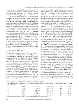
Table 1. Properties of cell-laden bio-inks ranging from 0 to 5 million cells/mL.
Cell concentration Viscosity (mPa.s) Surface tension (mN/m) Density (kg/m ) Nozzle radius (µm) Z value
3
(mil cells/mL)
0 0.687 72.12±0.47 1006.6±2.2 40 78.41
1.0 0.736 71.51±0.66 1007.3±2.6 40 72.92
2.0 0.776 66.52±0.90 1008.4±2.2 40 66.74
3.0 0.794 65.33±0.21 1009.2±2.5 40 64.67
4.0 0.828 63.48±0.82 1010.1±2.8 40 61.16
5.0 0.868 62.86±1.00 1012.0±2.2 40 58.11
Average viscosity at shear rate of 10,000 s , surface tension and density of the cell-laden bio-inks.
−1
28 International Journal of Bioprinting (2022)–Volume 8, Issue 1