
Page 69 - IJB-8-1
P. 69
Seiti, et al.
the two limits of the line (L and L right ), are retrieved. neglected, considering that both glass and Parylene-C
left
Furthermore, the overspray limits (O and O right ), have low thermal conductivity.
left
defined as the ones for which the grayscale intensity
reduces to 90% of the background, are computed 2.4. Biocompatibility and cellular adhesion
(Figure 2E). The average line and the overspray The biocompatibility of the NTE substrate has been proven
[27]
l ∑
( ) and
width are then calculated as w = k i= 1 wi by Ferraro et al. . Additional immunofluorescence
and cell viability assays were applied to test the
l
o ∑
w = k i= 1 wi l () L right ( ) i − L left ( ) i and biocompatibility and cellular adhesion on the Parylene-C
( ) , where wi =
o
w o ( ) i = O right ( ) i − O left ( ) i − wi coated NTE substrate and PEDOT: PSS samples.
( ) .
Specifically, immunofluorescence was executed to
l
2.3. Print technology transfer observe the cellular morphology and adhesion (nuclei and
In contrast to the typical empirical approaches applied to cytoskeleton) of human fibroblasts (HFs) and of human
iPSCs derived neural stem cells (NSCs) on the Parylene-
dedicated ink/substrate combinations, a methodology to C-coated NTE substrate, along with tests of NSCs on
allow direct transfer of printing strategies across different Matrigel-coated PEDOT: PSS samples. Cell viability
supports is proposed. It bases on the assumption Table 3. assays (direct and indirect) were instead executed to
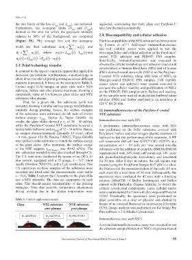
Contact angle (CA) images on glass slide and a NTE evaluate the cellular viability and proliferation of NSCs
substrate, before and after plasma treatment, showing a on the PEDOT: PSS samples only. Before cell seeding,
comparable value of CA between glass slides and NTE all the samples were washed in phosphate-buffered saline
substrates after plasma. solution (PBS) and further sterilized in an autoclave at
That, for a given ink, flat substrates (with low 121°C for 20 min.
porosity) showing a similar surface energy would behave
similarly during printing. Hence, the surface energies (1) Immunofluorescence of the Parylene-C-coated
of the substrates were estimated via ink tests of known NTE substrates
surface energy, γ TEST (Series A, Tigres GmbH). As
results, the glass slides showed a γ of 38 – 40 mN/m, Immunofluorescence with HFs
G
while the Parylene-C-coated NTE substrates revealed a A preliminary immunofluorescence assay with HFs
hydrophobic behavior and a γ NTE of 32 – 34 mN/m. Hence, was performed on the NTE substrates covered with
an oxygen plasma treatment (intensity 15 s/cm , offset Parylene-C before and after oxygen plasma treatment (3
2
~ 8 mm, power 150 W, Plasma T-SPOT, Tigres GmbH) replicas) to test the performance of the treatment. A HF
was applied on the substrates to match the surface energy cell suspension (BJ cell line ATCC CRL-2522™) at a
®
of the glass slides. After treatment, the surface energy concentration of 1 × 10 cells cm was poured onto the
5
–2
of the NTE support, γ NTE-Plasma , was 40-42 mN/m. The substrates with the addition of complete DMEM (DMEM
ink- substrates wettability was also checked through CA. supplemented with 10% fetal calf serum and 100 units/
The CA tests were conducted by means of an OCA 15 mL penicillin/streptomycin, Euroclone), and incubated
plus system, equipped with a 25 gauge, 1 – 1/2” blunt for 30 min. After 4 days in culture, the cell culture was
needle (Nordson 7018339), and a 2 μL sessile drop. Two secured using the Fix&Perm Sample Kit (SIC) to allow
®
CA repetitions on three samples of the substrates were the fixation and the permeabilization of the cells (15 min
recorded and fitted until the measurements were stable each step) for a total time of 30 min. Subsequently, the
(~ 10 s). Table 3 reports the CA results on the glass slide specimens were incubated for 45 min with a blocking
and a NTE substrate. The data are comparable to each solution (iBindTM ×5 Buffer, Invitrogen), and further
other. This should ensure transferability of the printing stained with Phalloidin (Sigma Aldrich), to detect the
strategies. Note that possible temperature phenomena cellular cytoskeletal components. Later, cellular nuclei
during printing due to the platen temperature were were counterstained for 5 min with the compound Hoechst
33342. Eventually, the specimens were positioned on
Table 3. Contact angle measurement glass coverslips on a drop of glycerol and studied by
Glass NTE substrate NTE substrate means of an inverted fluorescence microscope (Olympus
ante plasma post plasma IX70). Image analysis was performed via the Image-Pro
Plus software v.7.0 (Media Cybernetics).
Immunofluorescence with NSCs
A second immunofluorescence assay was executed to test
the adhesion and proliferation of NSCs on plasma-treated
International Journal of Bioprinting (2022)–Volume 8, Issue 1 55