
Page 107 - IJB-8-2
P. 107
Lee, et al.
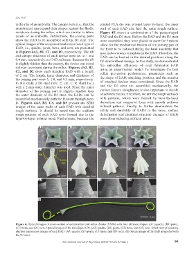
to the ribs of an umbrella. The canopy parts (i.e., flexible printed PLA die was printed layer-by-layer, the inner
membranes) can expand their shapes against the fluidic wall of each EAD also had the same rough surface.
resistance during the reflux, which are similar to fabric Figure 4E shows a combination of the penta-shaped
panels of an umbrella. Furthermore, the joining parts EAD and the DJ stent. Before the EAD and the DJ stent
allow the EAD to be assembled with the DJ stent. The were assembled, they were placed in water for 5 min to
optical images of the cross-sectional area of each type of allow for the mechanical friction of the joining part of
EAD, i.e., quadra, penta, hexa, and octa, are presented the EAD to be reduced during the hand assembly that
in Figures 4A1, B1, C1, and D1, respectively. The rib may induce cracks or rupture in the EAD. Therefore, the
and canopy thickness of each device were set to 1 and EAD can be located in the desired positions along the
0.4 mm, respectively, on CAD software. Because the rib DJ stent without damage. In this study, we demonstrated
is slightly thicker than the canopy, the device can avoid the anti-reflux efficiency of each fabricated EAD
rollover (overturn) during the reflux. Figures 4A2, B2, using an experimental model. To investigate the best
C2, and D2 show each standing EAD with a length reflux prevention performance, parameters such as
of 2 cm. The length, inner diameter, and thickness of
the joining part were 5, 1.8, and 0.4 mm, respectively. the shape of EAD, attaching position, and the number
In this study, a DJ stent (6Fr, 22 cm, C. R. Bard Inc.) of attached devices were considered. Since the EAD
with a 2-mm outer diameter was used. Since the inner and the DJ stent are assembled mechanically, the
diameter of the joining part is slightly smaller than surface feature (roughness) is also important to decide
the outer diameter of the DJ stent, the EADs can be attachment forces. Therefore, we utilized rough surfaces
assembled mechanically with the DJ stent through press with patterns, which were formed by layer-by-layer
fit. Figures 4A3, B3, C3, and D3 present the SEM deposition and compared them with smooth surfaces
images of the outer walls of each EAD with detailed without patterns. Finally, to further demonstrate the
rough surfaces. It should be noted that the uniform safety and durability of EADs in the urine, surface
rough patterns of each EAD were formed due to the deformation and chemical structure changes of EADs
layer-by-layer printed mold. Furthermore, because the were observed using artificial urine.
A1 B1 C1 D1
A2 B2 C2 D2
A3 B3 C3 D3
E
Figure 4. Optical images of cross-section of extraluminal anti-reflux diodes (EADs) with four different shapes: (A1) quadra, (B1) penta,
(C1) hexa, and (D1) octa. Optical images of the standing EADs: (A2) quadra, (B2) penta, (C2) hexa, and (D2) octa. Tilted view of scanning
electron microscopy images of each EAD: (A3) quadra, (B3) penta, (C3) hexa, and (D3) octa. (E) Optical image of the EAD integrated with
the DJ stent.
International Journal of Bioprinting (2022)–Volume 8, Issue 2 99