
Page 112 - IJB-8-2
P. 112
3D-Printing-Assisted EADs for Preventing VUR through DJ Stents
the DJ stent was pulled from the outside to the inside of of each artificial urine sample is presented in Table 2.
silicone rubber, as shown in Figure S3B2. Friction forces For the long-term experiment, the penta-shaped EADs
for each case were measured by pulling the DJ stent up were immersed in each artificial urine sample in a vial
along a displacement of 40 mm using a tensile testing and kept in a water bath at 36.5°C (body temperature)
machine. The experiments were conducted with pulling for 4 weeks. After 4 weeks, the EADs were washed
speeds of 50 and 100 mm/min, and repeated 3 times. with deionized (DI) water to investigate whether the
Figure 8D shows the averaged maximum load EAD was physically damaged or chemically corroded
between the EAD and inner wall of silicone rubber by urine. Figures 9A1, B1, C1, and D1 show the SEM
with respect to Positions A and B. Position A showed images of the EADs surface washed after immersion
maximum tensile load 116.65 and 96.04 mN at a pulling in low-pH, normal-pH, high-pH, and glucose urine,
speed of 50 and 100 mm/min, respectively. Position B respectively. No physical damage or solidified residues
showed maximum tensile load 157.74 and 263.34 mN of urine were observed after rinsing with DI water. This
at a pulling speed of 50 and 100 mm/min, respectively. characteristic is clearly distinct from the surfaces of the
Although the friction forces were present during the unwashed EADs. Figures 9A2, B2, C2, and D2 show
removal of DJ stent, these forces were considerably lower SEM images of the surfaces of unwashed EADs after
than the threshold force that can injure the inner wall of immersion in low-pH, normal-pH, high-pH, and glucose
the ureter (2.9 N) . The averaged maximum load at urine for 4 weeks, respectively. Each unwashed EAD
[50]
Position B showed higher values than those at Position was dried at 36.5°C for 12 h without being rinsed with
A, but still negligible compared to the threshold force of deionized water. The surfaces of the unwashed EADs
2.9 N. This was because more load was required to flip showed adhered urinary stones for EADs immersed in
the EAD at Position B, thus increasing the friction force low-pH, normal-pH, and high-pH urine, and solidified
simultaneously. However, it was noteworthy that once the glucose residues for the EAD immersed in glucose
EAD was flipped, the EAD was easily removed in the urine. These results imply that the washing process can
silicone rubber with lower friction forces. Consequently, remove residues of urine effectively, and thus the EADs
the fabricated EAD is expected not to damage or injure are reusable without physical damage after being washed
the inner wall (mucosa) of the ureter and urethra during with water.
both insertion and removal operations. To further demonstrate the chemically stable
structures of the EADs in urine, FTIR (Nicolet iS50,
3.3. Safety and durability test in urine Thermo Fisher Scientific Inc.) analysis was performed,
To investigate the safety and durability of EADs made as shown in Figure 10. The absorbance with respect
of Ecoflex (biocompatible) materials, the changes in to the wavenumber was measured to compare the
the surface and chemical structure of the EADs were chemical structures of the EADs before and after
thoroughly examined in four types of artificial urine immersion in artificial urine. For the bare EAD before
(Biozoa Biological Supply Co.): low-pH, normal-pH, immersion in urine, the absorbance bands were observed
high-pH, and glucose urine. The chemical composition at wavenumbers of 2963, 1260, 1089, 1018, and
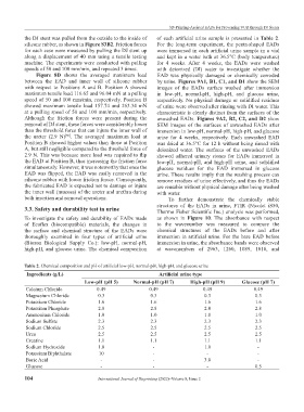
Table 2. Chemical composition and pH of artificial low-pH, normal-pH, high-pH, and glucose urine
Ingredients (g/L) Artificial urine type
Low-pH (pH 5) Normal-pH (pH 7) High-pH (pH 9) Glucose (pH 7)
Calcium Chloride 0.49 0.49 0.49 0.49
Magnesium Chloride 0.3 0.3 0.3 0.3
Potassium Chloride 1.6 1.6 1.6 1.6
Potassium Phosphate 2.8 2.8 2.8 2.8
Ammonium Chloride 1.0 1.0 1.0 1.0
Sodium Sulfate 2.3 2.3 2.3 2.3
Sodium Chloride 2.5 2.5 2.5 2.5
Urea 2.5 2.5 2.5 2.5
Creatine 1.1 1.1 1.1 1.1
Sodium Hydroxide 1.0 - 1.0 -
Potassium Biphthalate 10 - - -
Boric Acid - - 3.0 -
Glucose - - - 0.5
104 International Journal of Bioprinting (2022)–Volume 8, Issue 2