
Page 110 - IJB-8-2
P. 110
3D-Printing-Assisted EADs for Preventing VUR through DJ Stents
not present suitable voiding situations compared to this without patterns were prepared and evaluated to measure
study. Nonetheless, to demonstrate the use of the EAD in the adhesion forces.
the human body, further in vivo or clinical trial studies are Figure 8A shows the measured tensile load
needed. Although intraluminal reflux was not considered (i.e., adhesion force) of each specimen with respect
in this study, various intraluminal anti-reflux devices to the displacement at a pulling speed of 50 mm/min.
have been demonstrated and cone-shaped anti-reflux Consequently, the characteristics of surface types (with
devices are commercially available . When the EAD is and without patterns) were distinctly divided into “slip”
[15]
combined with these intraluminal anti-reflux devices, the and “miss” behaviors. For the joining part with rough
overall efficiency of reflux prevention alongside the stents patterns, the joining part was well attached on the surface
will be dramatically increased. Consequently, patients are of the DJ stent up to the pulling displacement of 20 –
expected to be free from the urological complications 25 mm, as confirmed from the linearly increased load.
caused by the VUR. When the pulling displacement exceeded 25 mm, the
measured load gradually decreased, meaning the joining
3.2. Mechanical adhesion and friction test part started slipping from the surface of the stent. In
It is important to avoid easy separation (disassembly) contrast, the joining part without rough patterns showed
of the EAD from the DJ stent during insertion into the a relatively low load and a sudden drop in pulling load
ureter line. Because of the features of the FDM-printing near a pulling displacement of 25 mm. This indicated
method, a rough surface with regular patterns appeared that the joining part was suddenly separated (missed)
both inside and outside the joining part, as shown in from the stent. The joining part without rough patterns
Figures 4A3, B3, C3, and D3. In this work, we utilized was also torn before separation from the DJ stent. Thus,
these unique patterns of the joining parts for stable and a smooth surface is more dangerous because the EAD
robust mechanical bonding with the DJ stent by enhancing can be broken away from the stent during insertion. In
the friction effect. To demonstrate the benefit of a rough summary, this result implies that the rough surface with
surface, the tensile (pulling) load at the interface between patterns formed by FDM printing exhibited relatively
the joining part and stent surface was measured using durable adhesion and longer attaching time with the DJ
a tensile testing machine (MCT-2150, A&D Co.). To stent than the smooth surface without patterns.
investigate the effect of the patterned surface, two types Figure 8B shows the average maximum load
of specimens were prepared, that is, with and without for the joining parts with rough and smooth surfaces.
patterned rough surfaces of the joining part; these Consequently, specimens with and without surface
specimens had a length, inner diameter, and thickness of 5, patterns showed maximum tensile loads of 1274.4 and
1.8, and 0.4 mm, respectively. Casting fabrication methods 1005.6 mN, respectively. The maximum average load of
(Figure 3) were also used in fabricating the specimens with the rough surface with patterns was 26.7% higher than
and without a rough surface. In this study, an acrylonitrile that of the smooth surface without patterns. A higher
butadiene styrene (ABS) filament was used to create the maximum load might be caused by the water (or urine)
joining part without patterns. Since ABS melts in acetone, between the grooved patterns and the surface of the
the rough surface of the ABS die was polished by acetone stent. The frictional force increased with the contact area
while maintaining the same diameter of the PLA die when the same normal pressure was applied. Therefore,
(Figure S1 in Supplementary File). After assembly with the applied load of a specimen with a smooth surface
the DJ stent, adhesion forces were measured by holding was assumed to be larger than that of a rough surface.
the DJ stent and pulling it at a speed of 50 mm/min and up However, if water is present between the smooth surface
to a displacement of 40 mm using a tensile testing machine and the surface of the DJ stent, the actual contact area at
(Figure S2 in Supplementary File). In this experiment, the interface significantly decreases due to the thin liquid
three different specimens for each joining part with and film, thus considerably reducing the frictional stress. In
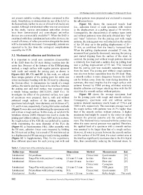
Table 1. Comparison of the anti-reflux devices
Reflux type Material Dimensions Method Measurement Ref.
Intraluminal Polyurethane N/A Clinical trial Questionnaire [15]
Intraluminal Tango Plus with parylene C coating 2.8 mm×5.3 mm In vitro Flow rate [21]
Intraluminal Silicone sleeve 15 mm×26 mm In vitro/ Flow rate/ [23]
Clinical trial Cystogram
Intraluminal Tango Plus with parylene C coating 2.8 mm×5.3 mm In vivo VUR grade [25]
Intraluminal Polyurethane with hydrogel coating N/A Clinical trial Questionnaire [27]
Extraluminal Ecoflex 10 mm × 20 mm In vitro Reflux height This work
102 International Journal of Bioprinting (2022)–Volume 8, Issue 2