
Page 113 - IJB-8-2
P. 113
Lee, et al.
A1 B1 C1 D1
A2 B2 C2 D2
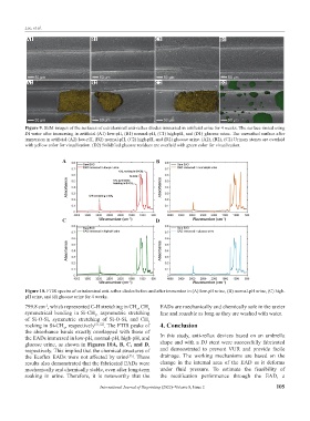
Figure 9. SEM images of the surfaces of extraluminal anti-reflux diodes immersed in artificial urine for 4 weeks. The surface rinsed using
DI water after immersing in artificial (A1) low-pH, (B1) normal-pH, (C1) high-pH, and (D1) glucose urine. The unwashed surface after
immersion in artificial (A2) low-pH, (B2) normal-pH, (C2) high-pH, and (D2) glucose urine. (A2), (B2), (C2) Urinary stones are overlaid
with yellow color for visualization. (D2) Solidified glucose residues are overlaid with green color for visualization.
A B
C D
Figure 10. FTIR spectra of extraluminal anti-reflux diodes before and after immersion in (A) low-pH urine, (B) normal-pH urine, (C) high-
pH urine, and (d) glucose urine for 4 weeks.
799.8 cm , which represented C-H stretching in CH , CH EADs are mechanically and chemically safe in the ureter
-1
3
3
symmetrical bending in Si-CH , asymmetric stretching line and reusable as long as they are washed with water.
3
of Si-O-Si, symmetric stretching of Si-O-Si, and CH
3
rocking in Si-CH , respectively [51,52] . The FTIR peaks of 4. Conclusion
3
the absorbance bands exactly overlapped with those of
the EADs immersed in low-pH, normal-pH, high-pH, and In this study, anti-reflux devices based on an umbrella
glucose urine, as shown in Figures 10A, B, C, and D, shape and with a DJ stent were successfully fabricated
respectively. This implied that the chemical structures of and demonstrated to prevent VUR and provide facile
the Ecoflex EADs were not affected by urine . These drainage. The working mechanisms are based on the
[53]
results also demonstrated that the fabricated EADs were change in the internal area of the EAD as it deforms
mechanically and chemically stable, even after long-term under fluid pressure. To estimate the feasibility of
soaking in urine. Therefore, it is noteworthy that the the rectification performance through the EAD, a
International Journal of Bioprinting (2022)–Volume 8, Issue 2 105