
Page 127 - IJB-8-2
P. 127
Dee, et al.
A B on heated gypsum (Figure 8A). At 30% infill density,
filaments were widely spaced apart, which is expected to
be favorable for vascularization (Figure 8B, left). In the
second example, a higher infill density of 50% was used
to print a six-layer thick portion of a bone plate at room
temperature (Figure 8B, right). The outer surface finish
appeared rough, as further discussed in Supplementary
C File: Section: 4.2. Post-processing would be required to
improve the surface finish although surface roughness
could promote cell attachment and bone growth . Micro-
[7]
porosity could be further explored using sacrificial organic
material or foaming agents, for example. The effects of
the scaffold microstructure and surface roughness on
osteoinductivity, cell differentiation and proliferation, and
D bone remodeling remains to be explored.
Furthermore, our approach also permitted the
printing of overhanging filaments that support their own
weight. In our buildability test modified from Ribeiro et al.
(2018) , underlying supports of three layers tall were
[41]
extruded at spacing’s ranging from 1.0 mm to 3.0 mm
(Figure 8C). The fourth and topmost layer of filaments
spanned across the supports below. Filaments extruded
through 0.58 mm nozzle at f = 500% could be continuously
extruded across supports up to 2.8 mm apart reliably. The
topmost layer was able to support the weight of additional
filaments without collapse and the filaments did not show
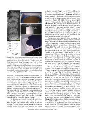
Figure 7. (A) Electron micrograph of the cross-section of a 5-layer deformation (see Figure S6 in Supplementary File).
print, scale bar: 200 µm. Height of calcined parts, printed at room Besides, the rectangular shape of majority of the pores as
temperature, d = 0.58 mm, f = 500%, v = 5 mm/s. Dashed line
shows linear fit. (B) Optical photograph of a 6 layer-print after seen from the top view also points to good print accuracy
[42]
debinding, placed next to a Singapore 5-cent coin 16.75 mm in and print fidelity . 24 vol% brushite ink with higher
[43]
diameter. (C) Optical photograph of a 20-layer brushite cylindrical storage modulus is expected to bridge wider spans .
shell printed on heated gypsum. Background contains a stack of ten For a 0.58 mm nozzle using flow rate of 500%,
microscope glass slides 10.25 mm tall. (D) 10-layer brushite jigsaw filament fusion occurred for center-to-center spacing of
piece printed on heated gypsum (left). 5 mm thick jigsaw piece 1.0 mm (Figure 8C). The post-calcination filament width
printed from PLA by FDM (right). Ruler in centimeters. is 0.92 ± 0.03 mm. The horizontal print resolution is
limited by the filament thickness after extrusion, which
processes . Implantation in sheep tibiae found that the has a minimum of 0.7 mm in the green body for a nozzle
[39]
addition of 28 wt% CPP to brushite bone cement promoted diameter of 0.41 mm and flow rate of 400%. Although
bone mineralization in vivo as osteoblasts hydrolyzed line-by-line robocasting is generally not well-suited for
pyrophosphate into orthophosphate ions . Better printing dense monolithic structures due to voids between
[40]
osseointegration of bioceramics with pyrophosphate and printed filaments [30,36,44] , we could make use of filament
bone mineralization in humans have also been clinically fusion to minimize porosity in bulk structures by careful
reported . Prolonged exposure to pyrophosphate was tuning of the infill density I . At infill density I = 30%,
[39]
f
f
found to stimulate osteoblast differentiation in vitro . there was no overlap between adjacent filaments. At
[39]
The bioactivity of β-CPP makes it a potential material in I ≥ 70%, the overlap between adjacent filaments
f
applications such as porous bone scaffolds, bone screws, caused the layer height to increase unevenly, leading to
bone coatings or bone plates. Osseointegration of CaP- deviation from the print design (Figure 8D and E). Infill
based bone plates could potentially eliminate the need for density ~50% could be chosen to print bulk structures
surgical removal of metallic internal fixation. with minimal porosity and good interfilament interface
To demonstrate various biomedical applications (Figure 8F), It is also notable that the calcination step of
requiring different macro-porosity, multi-layered structures the multi-layered bulk parts did not cause defects such
were designed with different infill density. In the first as microcracks or delamination due to the consolidation
example, a 16 mm diameter scaffold resembling commonly without shrinkage. Anisotropic ceramic materials present
robocast porous cancellous bone scaffolds was printed a tendency to exhibit strong anisotropic shrinkage. This is
International Journal of Bioprinting (2022)–Volume 8, Issue 2 119