
Page 147 - IJB-8-2
P. 147
Kanaki, et al.
A
B
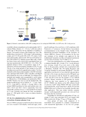
Figure 2. Schematic representation of the LIFT printing process for coating the PMMA MNs. (A) LIFT setup. (B) Coating process.
controlled column compartment, and an autosampler. A dC18 specific pathogen-free conditions, in full compliance with
column (Water, Atlantis, 2.1 × 50 mm, 3 μM) was used at Federation of Laboratory Animal Science Associations
a flow rate of 300 μl/min for the separation of analytes of recommendations in the Animal House Facility of the
interest. The injection volume of the samples was 10 μl. The Biomedical Research Foundation of the Academy of
mobile phase consisted of A: 100% water, 2 mM ammonium Athens (BRFAA, Greece). All procedures for the care
acetate, and 0.1% FA and B: 90% ACN, 10% water, 2 mM and treatment of the animals were approved by the
ammonium acetate, and 0.1% FA. MS was performed on an Institutional Committee on Ethics of Animal Experiments
API 5500 QTRAP LC-MS/MS system fitted with a Turbo and the Attica Prefecture. No:971840/16-11-21.
Ion Spray source and a hybrid triple quadrupole/linear ion Gem was administered to mice either transdermally
trap mass spectrometer (AB SCIEX LLC, CA, USA). The or intraperitoneally (IP). A total of 36, 5-week-old male
standard solutions within a concentration range of 10 – mice (average weight 20 g) were used in this study.
200 ng/mL of Gem were prepared. Samples of 100, 375, and Prior to the procedure, animals were anesthetized with
750 μg of the initial amount of Gem printed by the LIFT a Ketamine: Xylazine mix (90 mg/kg Ketamine and
technique were used for the quantification studies. The Gem 10 mg/kg Xylazine) as previously described . For MN
[35]
standards and the laser printed samples on the MN substrates application onto the skin of mice, the left side just above
were extracted with MeOH (100%) and then centrifuged the flank of the mouse was shaved and the MN patch was
with a Speed Vac for 5 min at 16000 rpm. The obtained Gem placed and pressed with the thumb for approximately
standards and samples were resuspended in mobile phase 2 min and left on the skin for another 10 min without
A, followed by a 10-fold dilution, and analyzed by HPLC- pressure. Manual insertion of MN patches by human
MS/MS. Warfarin was used as internal standard (IS). The volunteers has been reported to be approximately
gradient methodology was as follows: 0 – 1.5 min: 100% 20 N . PMMA was chosen as the MN polymer based
[4]
A, 1.5 – 5 min: 40% A – 60% B, 5 – 10 min: 100% A. The on previous work using PMMA MN tips coated with
[6]
primary MRM transitions for Gem and warfarin were: m/z water-dissolvable, drug-loaded layer which showed that
264.1→112.0 and 309.1→162.9, respectively. Gem was PMMA MNs can be removed successfully from the skin
eluted with a retention time of 1.6 min and the IS was eluted after application. This may reduce toxicity concerns
at 5.2 min. Linearity was shown for Gem concentration because nonbiodegradable polymers are removed from
range of 1–500 ng/mL with a lower on-column detection the tissue after the treatment.
limit of 10 pg. Regarding the transdermal application of Gem, six
2.9. In vivo administration of Gem and groups of mice were generated (n≥3) and treated with
extraction of mouse plasma Gem MNs loaded with 100 μg, 375 μg, or 750 μg of Gem.
Four groups of mice, each composed of 4
Mice (C57Bl/6) were purchased from Jackson laboratories individuals were generated to compare intraperitoneal
and were housed in individually ventilated cages, under with transdermal application. In the first two groups,
International Journal of Bioprinting (2022)–Volume 8, Issue 3 139